Zinc »
PDB 3woj-3x0d »
3wus »
Zinc in PDB 3wus: Crystal Structure of the Vif-Binding Domain of Human APOBEC3F
Protein crystallography data
The structure of Crystal Structure of the Vif-Binding Domain of Human APOBEC3F, PDB code: 3wus
was solved by
M.Nakashima,
T.Kawamura,
H.Ode,
N.Watanabe,
Y.Iwatani,
with X-Ray Crystallography technique. A brief refinement statistics is given in the table below:
| Resolution Low / High (Å) | 50.00 / 2.54 |
| Space group | P 31 2 1 |
| Cell size a, b, c (Å), α, β, γ (°) | 117.294, 117.294, 78.960, 90.00, 90.00, 120.00 |
| R / Rfree (%) | 20 / 25.6 |
Zinc Binding Sites:
The binding sites of Zinc atom in the Crystal Structure of the Vif-Binding Domain of Human APOBEC3F
(pdb code 3wus). This binding sites where shown within
5.0 Angstroms radius around Zinc atom.
In total 2 binding sites of Zinc where determined in the Crystal Structure of the Vif-Binding Domain of Human APOBEC3F, PDB code: 3wus:
Jump to Zinc binding site number: 1; 2;
In total 2 binding sites of Zinc where determined in the Crystal Structure of the Vif-Binding Domain of Human APOBEC3F, PDB code: 3wus:
Jump to Zinc binding site number: 1; 2;
Zinc binding site 1 out of 2 in 3wus
Go back to
Zinc binding site 1 out
of 2 in the Crystal Structure of the Vif-Binding Domain of Human APOBEC3F
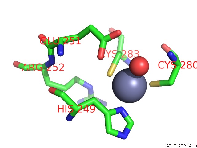
Mono view
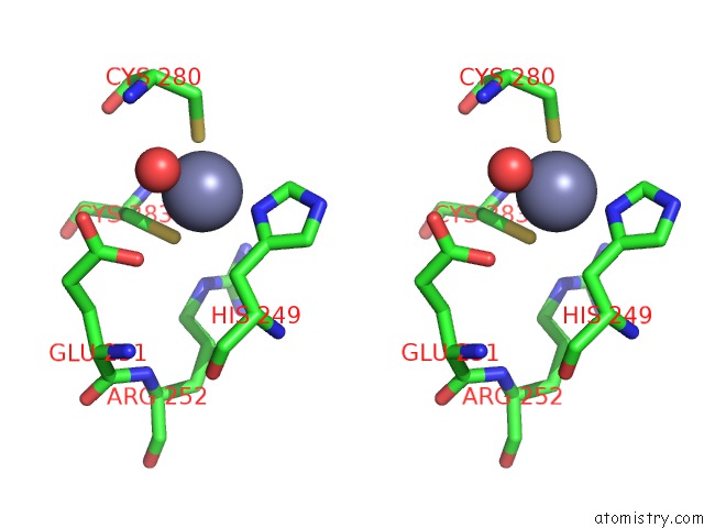
Stereo pair view
Mono view
Stereo pair view
A full contact list of Zinc with other atoms in the Zn binding
site number 1 of Crystal Structure of the Vif-Binding Domain of Human APOBEC3F within 5.0Å range:
|
Zinc binding site 2 out of 2 in 3wus
Go back to
Zinc binding site 2 out
of 2 in the Crystal Structure of the Vif-Binding Domain of Human APOBEC3F
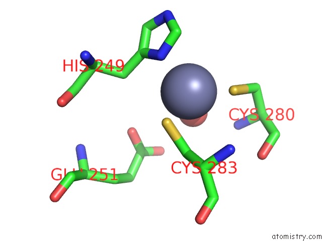
Mono view
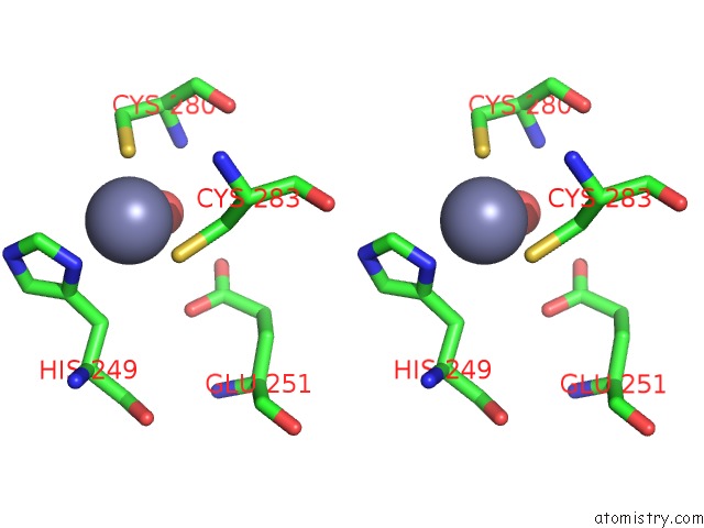
Stereo pair view
Mono view
Stereo pair view
A full contact list of Zinc with other atoms in the Zn binding
site number 2 of Crystal Structure of the Vif-Binding Domain of Human APOBEC3F within 5.0Å range:
|
Reference:
M.Nakashima,
H.Ode,
T.Kawamura,
S.Kitamura,
Y.Naganawa,
H.Awazu,
S.Tsuzuki,
K.Matsuoka,
M.Nemoto,
A.Hachiya,
W.Sugiura,
Y.Yokomaku,
N.Watanabe,
Y.Iwatani.
Structural Insights Into Hiv-1 Vif-APOBEC3F Interaction. J.Virol. V. 90 1034 2015.
ISSN: ISSN 0022-538X
PubMed: 26537685
DOI: 10.1128/JVI.02369-15
Page generated: Sat Oct 26 18:19:11 2024
ISSN: ISSN 0022-538X
PubMed: 26537685
DOI: 10.1128/JVI.02369-15
Last articles
Zn in 9MJ5Zn in 9HNW
Zn in 9G0L
Zn in 9FNE
Zn in 9DZN
Zn in 9E0I
Zn in 9D32
Zn in 9DAK
Zn in 8ZXC
Zn in 8ZUF