Zinc »
PDB 3uk4-3v38 »
3ux3 »
Zinc in PDB 3ux3: Crystal Structure of Domain-Swapped FAM96A Minor Dimer
Protein crystallography data
The structure of Crystal Structure of Domain-Swapped FAM96A Minor Dimer, PDB code: 3ux3
was solved by
K.-E.Chen,
B.Kobe,
J.L.Martin,
with X-Ray Crystallography technique. A brief refinement statistics is given in the table below:
| Resolution Low / High (Å) | 34.24 / 1.80 |
| Space group | P 1 21 1 |
| Cell size a, b, c (Å), α, β, γ (°) | 49.179, 52.246, 65.893, 90.00, 104.56, 90.00 |
| R / Rfree (%) | 20 / 24.6 |
Zinc Binding Sites:
The binding sites of Zinc atom in the Crystal Structure of Domain-Swapped FAM96A Minor Dimer
(pdb code 3ux3). This binding sites where shown within
5.0 Angstroms radius around Zinc atom.
In total 3 binding sites of Zinc where determined in the Crystal Structure of Domain-Swapped FAM96A Minor Dimer, PDB code: 3ux3:
Jump to Zinc binding site number: 1; 2; 3;
In total 3 binding sites of Zinc where determined in the Crystal Structure of Domain-Swapped FAM96A Minor Dimer, PDB code: 3ux3:
Jump to Zinc binding site number: 1; 2; 3;
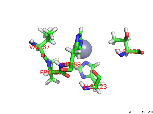
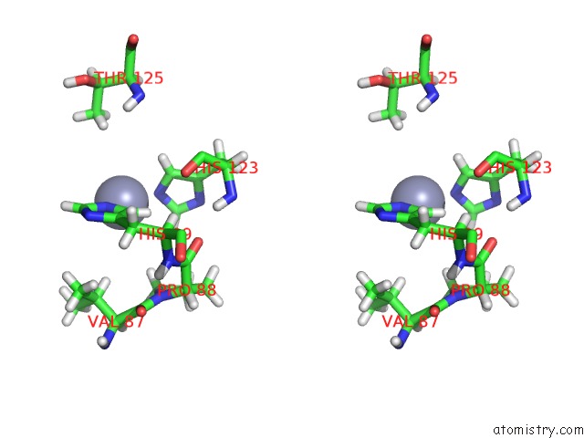
Zinc binding site 1 out of 3 in 3ux3
Go back to
Zinc binding site 1 out
of 3 in the Crystal Structure of Domain-Swapped FAM96A Minor Dimer
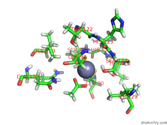
Mono view
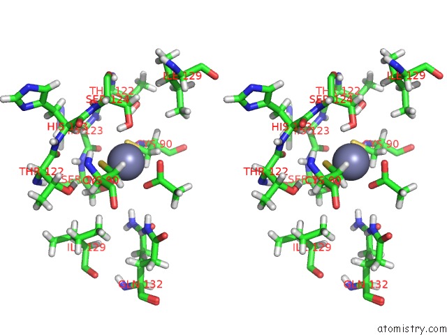
Stereo pair view
Mono view
Stereo pair view
A full contact list of Zinc with other atoms in the Zn binding
site number 1 of Crystal Structure of Domain-Swapped FAM96A Minor Dimer within 5.0Å range:
|
Zinc binding site 2 out of 3 in 3ux3
Go back to
Zinc binding site 2 out
of 3 in the Crystal Structure of Domain-Swapped FAM96A Minor Dimer
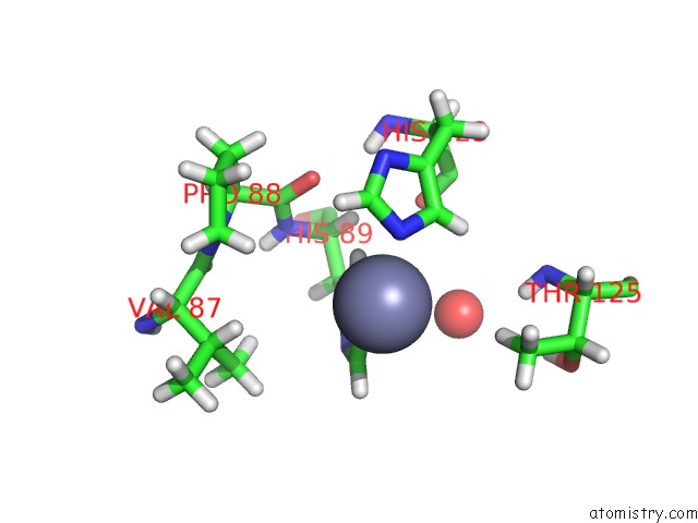
Mono view
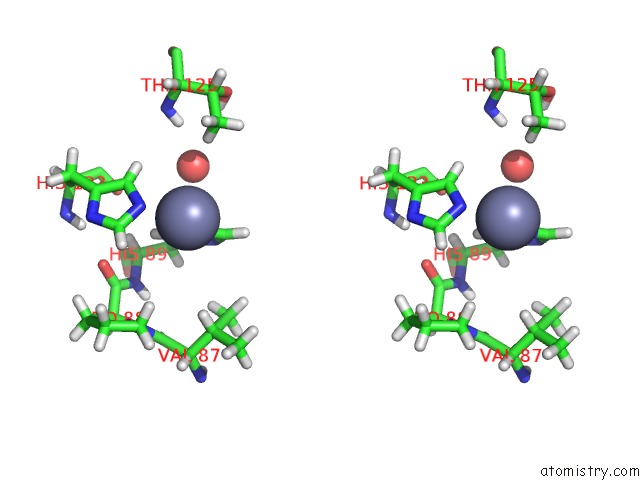
Stereo pair view
Mono view
Stereo pair view
A full contact list of Zinc with other atoms in the Zn binding
site number 2 of Crystal Structure of Domain-Swapped FAM96A Minor Dimer within 5.0Å range:
|
Zinc binding site 3 out of 3 in 3ux3
Go back to
Zinc binding site 3 out
of 3 in the Crystal Structure of Domain-Swapped FAM96A Minor Dimer
Mono view
Stereo pair view
Mono view
Stereo pair view
A full contact list of Zinc with other atoms in the Zn binding
site number 3 of Crystal Structure of Domain-Swapped FAM96A Minor Dimer within 5.0Å range:
|
Reference:
K.E.Chen,
A.A.Richards,
J.K.Ariffin,
I.L.Ross,
M.J.Sweet,
S.Kellie,
B.Kobe,
J.L.Martin.
The Mammalian DUF59 Protein FAM96A Forms Two Distinct Types of Domain-Swapped Dimer. Acta Crystallogr.,Sect.D V. 68 637 2012.
ISSN: ESSN 1399-0047
PubMed: 22683786
DOI: 10.1107/S0907444912006592
Page generated: Sat Oct 26 17:29:54 2024
ISSN: ESSN 1399-0047
PubMed: 22683786
DOI: 10.1107/S0907444912006592
Last articles
Zn in 9MJ5Zn in 9HNW
Zn in 9G0L
Zn in 9FNE
Zn in 9DZN
Zn in 9E0I
Zn in 9D32
Zn in 9DAK
Zn in 8ZXC
Zn in 8ZUF