Zinc »
PDB 3u6q-3uec »
3u9g »
Zinc in PDB 3u9g: Crystal Structure of the Zinc Finger Antiviral Protein
Protein crystallography data
The structure of Crystal Structure of the Zinc Finger Antiviral Protein, PDB code: 3u9g
was solved by
S.Chen,
Y.Xu,
K.Zhang,
X.Wang,
J.Sun,
G.Gao,
Y.Liu,
with X-Ray Crystallography technique. A brief refinement statistics is given in the table below:
| Resolution Low / High (Å) | 32.49 / 1.80 |
| Space group | P 32 2 1 |
| Cell size a, b, c (Å), α, β, γ (°) | 52.894, 52.894, 138.275, 90.00, 90.00, 120.00 |
| R / Rfree (%) | 19.1 / 22.8 |
Zinc Binding Sites:
The binding sites of Zinc atom in the Crystal Structure of the Zinc Finger Antiviral Protein
(pdb code 3u9g). This binding sites where shown within
5.0 Angstroms radius around Zinc atom.
In total 4 binding sites of Zinc where determined in the Crystal Structure of the Zinc Finger Antiviral Protein, PDB code: 3u9g:
Jump to Zinc binding site number: 1; 2; 3; 4;
In total 4 binding sites of Zinc where determined in the Crystal Structure of the Zinc Finger Antiviral Protein, PDB code: 3u9g:
Jump to Zinc binding site number: 1; 2; 3; 4;
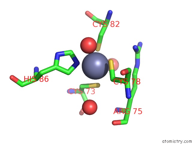
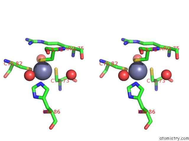
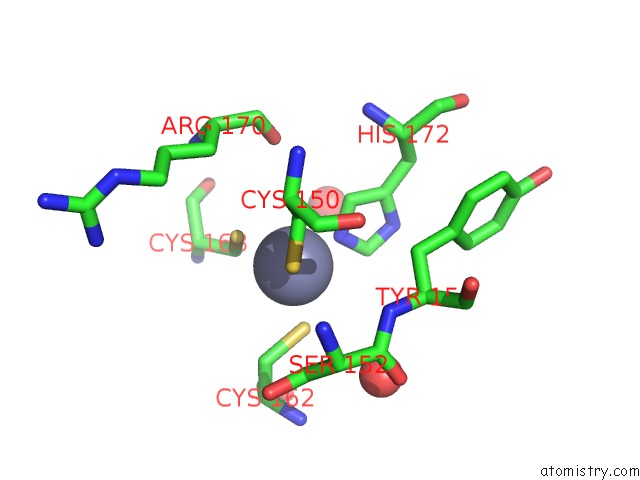
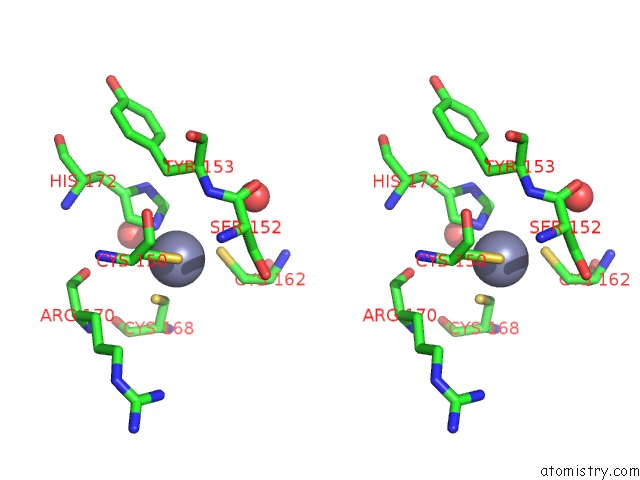
Zinc binding site 1 out of 4 in 3u9g
Go back to
Zinc binding site 1 out
of 4 in the Crystal Structure of the Zinc Finger Antiviral Protein
Mono view
Stereo pair view
Mono view
Stereo pair view
A full contact list of Zinc with other atoms in the Zn binding
site number 1 of Crystal Structure of the Zinc Finger Antiviral Protein within 5.0Å range:
|
Zinc binding site 2 out of 4 in 3u9g
Go back to
Zinc binding site 2 out
of 4 in the Crystal Structure of the Zinc Finger Antiviral Protein
Mono view
Stereo pair view
Mono view
Stereo pair view
A full contact list of Zinc with other atoms in the Zn binding
site number 2 of Crystal Structure of the Zinc Finger Antiviral Protein within 5.0Å range:
|
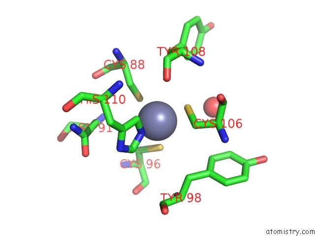
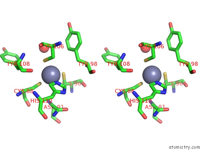
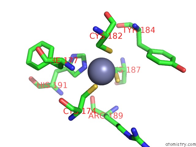
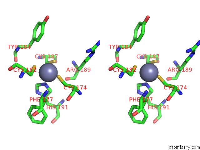
Zinc binding site 3 out of 4 in 3u9g
Go back to
Zinc binding site 3 out
of 4 in the Crystal Structure of the Zinc Finger Antiviral Protein
Mono view
Stereo pair view
Mono view
Stereo pair view
A full contact list of Zinc with other atoms in the Zn binding
site number 3 of Crystal Structure of the Zinc Finger Antiviral Protein within 5.0Å range:
|
Zinc binding site 4 out of 4 in 3u9g
Go back to
Zinc binding site 4 out
of 4 in the Crystal Structure of the Zinc Finger Antiviral Protein
Mono view
Stereo pair view
Mono view
Stereo pair view
A full contact list of Zinc with other atoms in the Zn binding
site number 4 of Crystal Structure of the Zinc Finger Antiviral Protein within 5.0Å range:
|
Reference:
S.Chen,
Y.Xu,
K.Zhang,
X.Wang,
J.Sun,
G.Gao,
Y.Liu.
Structure of N-Terminal Domain of Zap Indicates How A Zinc-Finger Protein Recognizes Complex Rna. Nat.Struct.Mol.Biol. V. 19 430 2012.
ISSN: ISSN 1545-9993
PubMed: 22407013
DOI: 10.1038/NSMB.2243
Page generated: Sat Oct 26 17:03:57 2024
ISSN: ISSN 1545-9993
PubMed: 22407013
DOI: 10.1038/NSMB.2243
Last articles
Zn in 9MJ5Zn in 9HNW
Zn in 9G0L
Zn in 9FNE
Zn in 9DZN
Zn in 9E0I
Zn in 9D32
Zn in 9DAK
Zn in 8ZXC
Zn in 8ZUF