Zinc »
PDB 3tus-3u6p »
3u52 »
Zinc in PDB 3u52: X-Ray Crystal Structure of Xenon-Pressurized Phenol Hydroxylase From Pseudomonas Sp. OX1
Protein crystallography data
The structure of X-Ray Crystal Structure of Xenon-Pressurized Phenol Hydroxylase From Pseudomonas Sp. OX1, PDB code: 3u52
was solved by
M.S.Mccormick,
S.J.Lippard,
with X-Ray Crystallography technique. A brief refinement statistics is given in the table below:
| Resolution Low / High (Å) | 38.00 / 1.95 |
| Space group | P 21 21 21 |
| Cell size a, b, c (Å), α, β, γ (°) | 83.942, 141.761, 181.207, 90.00, 90.00, 90.00 |
| R / Rfree (%) | 18.3 / 22.8 |
Other elements in 3u52:
The structure of X-Ray Crystal Structure of Xenon-Pressurized Phenol Hydroxylase From Pseudomonas Sp. OX1 also contains other interesting chemical elements:
| Xenon | (Xe) | 24 atoms |
| Iron | (Fe) | 4 atoms |
| Copper | (Cu) | 2 atoms |
Zinc Binding Sites:
The binding sites of Zinc atom in the X-Ray Crystal Structure of Xenon-Pressurized Phenol Hydroxylase From Pseudomonas Sp. OX1
(pdb code 3u52). This binding sites where shown within
5.0 Angstroms radius around Zinc atom.
In total 2 binding sites of Zinc where determined in the X-Ray Crystal Structure of Xenon-Pressurized Phenol Hydroxylase From Pseudomonas Sp. OX1, PDB code: 3u52:
Jump to Zinc binding site number: 1; 2;
In total 2 binding sites of Zinc where determined in the X-Ray Crystal Structure of Xenon-Pressurized Phenol Hydroxylase From Pseudomonas Sp. OX1, PDB code: 3u52:
Jump to Zinc binding site number: 1; 2;
Zinc binding site 1 out of 2 in 3u52
Go back to
Zinc binding site 1 out
of 2 in the X-Ray Crystal Structure of Xenon-Pressurized Phenol Hydroxylase From Pseudomonas Sp. OX1
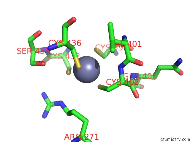
Mono view
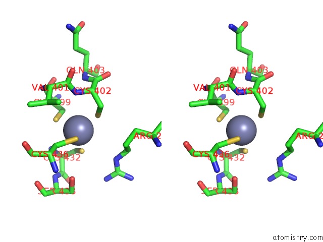
Stereo pair view
Mono view
Stereo pair view
A full contact list of Zinc with other atoms in the Zn binding
site number 1 of X-Ray Crystal Structure of Xenon-Pressurized Phenol Hydroxylase From Pseudomonas Sp. OX1 within 5.0Å range:
|
Zinc binding site 2 out of 2 in 3u52
Go back to
Zinc binding site 2 out
of 2 in the X-Ray Crystal Structure of Xenon-Pressurized Phenol Hydroxylase From Pseudomonas Sp. OX1
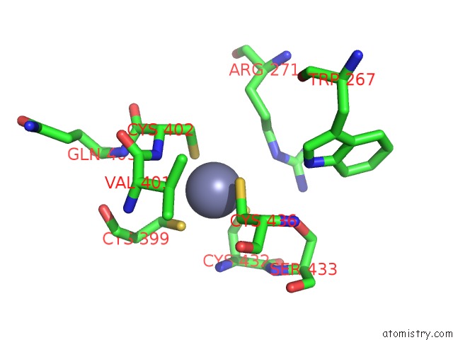
Mono view
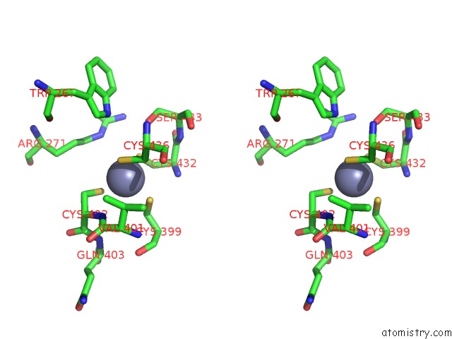
Stereo pair view
Mono view
Stereo pair view
A full contact list of Zinc with other atoms in the Zn binding
site number 2 of X-Ray Crystal Structure of Xenon-Pressurized Phenol Hydroxylase From Pseudomonas Sp. OX1 within 5.0Å range:
|
Reference:
M.S.Mccormick,
S.J.Lippard.
Analysis of Substrate Access to Active Sites in Bacterial Multicomponent Monooxygenase Hydroxylases: X-Ray Crystal Structure of Xenon-Pressurized Phenol Hydroxylase From Pseudomonas Sp. OX1. Biochemistry V. 50 11058 2011.
ISSN: ISSN 0006-2960
PubMed: 22136180
DOI: 10.1021/BI201248B
Page generated: Sat Oct 26 16:54:07 2024
ISSN: ISSN 0006-2960
PubMed: 22136180
DOI: 10.1021/BI201248B
Last articles
Zn in 9MJ5Zn in 9HNW
Zn in 9G0L
Zn in 9FNE
Zn in 9DZN
Zn in 9E0I
Zn in 9D32
Zn in 9DAK
Zn in 8ZXC
Zn in 8ZUF