Zinc »
PDB 3tge-3tus »
3tsq »
Zinc in PDB 3tsq: Crystal Structure of E. Coli Hypf with Atp and Carbamoyl Phosphate
Protein crystallography data
The structure of Crystal Structure of E. Coli Hypf with Atp and Carbamoyl Phosphate, PDB code: 3tsq
was solved by
S.Petkun,
R.Shi,
Y.Li,
M.Cygler,
with X-Ray Crystallography technique. A brief refinement statistics is given in the table below:
| Resolution Low / High (Å) | 100.41 / 2.40 |
| Space group | P 21 21 21 |
| Cell size a, b, c (Å), α, β, γ (°) | 46.530, 78.287, 200.815, 90.00, 90.00, 90.00 |
| R / Rfree (%) | 15.4 / 23.3 |
Other elements in 3tsq:
The structure of Crystal Structure of E. Coli Hypf with Atp and Carbamoyl Phosphate also contains other interesting chemical elements:
| Magnesium | (Mg) | 1 atom |
Zinc Binding Sites:
The binding sites of Zinc atom in the Crystal Structure of E. Coli Hypf with Atp and Carbamoyl Phosphate
(pdb code 3tsq). This binding sites where shown within
5.0 Angstroms radius around Zinc atom.
In total 3 binding sites of Zinc where determined in the Crystal Structure of E. Coli Hypf with Atp and Carbamoyl Phosphate, PDB code: 3tsq:
Jump to Zinc binding site number: 1; 2; 3;
In total 3 binding sites of Zinc where determined in the Crystal Structure of E. Coli Hypf with Atp and Carbamoyl Phosphate, PDB code: 3tsq:
Jump to Zinc binding site number: 1; 2; 3;
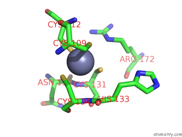
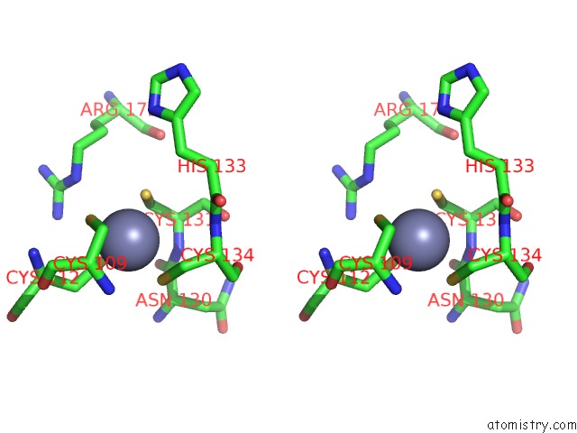
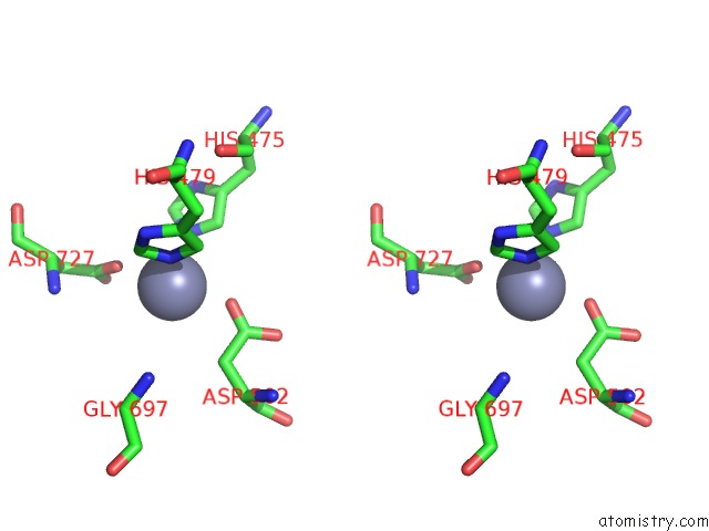
Zinc binding site 1 out of 3 in 3tsq
Go back to
Zinc binding site 1 out
of 3 in the Crystal Structure of E. Coli Hypf with Atp and Carbamoyl Phosphate
Mono view
Stereo pair view
Mono view
Stereo pair view
A full contact list of Zinc with other atoms in the Zn binding
site number 1 of Crystal Structure of E. Coli Hypf with Atp and Carbamoyl Phosphate within 5.0Å range:
|
Zinc binding site 2 out of 3 in 3tsq
Go back to
Zinc binding site 2 out
of 3 in the Crystal Structure of E. Coli Hypf with Atp and Carbamoyl Phosphate
Mono view
Stereo pair view
Mono view
Stereo pair view
A full contact list of Zinc with other atoms in the Zn binding
site number 2 of Crystal Structure of E. Coli Hypf with Atp and Carbamoyl Phosphate within 5.0Å range:
|
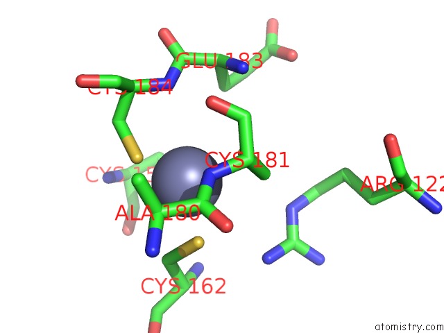
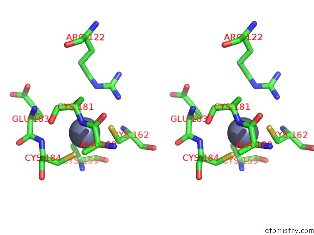
Zinc binding site 3 out of 3 in 3tsq
Go back to
Zinc binding site 3 out
of 3 in the Crystal Structure of E. Coli Hypf with Atp and Carbamoyl Phosphate

Mono view
Stereo pair view
Mono view
Stereo pair view
A full contact list of Zinc with other atoms in the Zn binding
site number 3 of Crystal Structure of E. Coli Hypf with Atp and Carbamoyl Phosphate within 5.0Å range:
|
Reference:
S.Petkun,
R.Shi,
Y.Li,
A.Asinas,
C.Munger,
L.Zhang,
M.Waclawek,
B.Soboh,
R.G.Sawers,
M.Cygler.
Structure of Hydrogenase Maturation Protein Hypf with Reaction Intermediates Shows Two Active Sites. Structure V. 19 1773 2011.
ISSN: ISSN 0969-2126
PubMed: 22153500
DOI: 10.1016/J.STR.2011.09.023
Page generated: Wed Aug 20 14:33:18 2025
ISSN: ISSN 0969-2126
PubMed: 22153500
DOI: 10.1016/J.STR.2011.09.023
Last articles
Zn in 4KJMZn in 4KJV
Zn in 4KJU
Zn in 4KIS
Zn in 4KJG
Zn in 4KFZ
Zn in 4KH1
Zn in 4KH0
Zn in 4KGZ
Zn in 4KGX