Zinc »
PDB 3tg5-3tty »
3tn2 »
Zinc in PDB 3tn2: Structure Analysis of MIP1-Beta P8A
Protein crystallography data
The structure of Structure Analysis of MIP1-Beta P8A, PDB code: 3tn2
was solved by
Q.Guo,
W.J.Tang,
with X-Ray Crystallography technique. A brief refinement statistics is given in the table below:
| Resolution Low / High (Å) | 29.45 / 1.60 |
| Space group | C 1 2 1 |
| Cell size a, b, c (Å), α, β, γ (°) | 51.062, 36.972, 31.061, 90.00, 108.54, 90.00 |
| R / Rfree (%) | 15.7 / 19.9 |
Zinc Binding Sites:
The binding sites of Zinc atom in the Structure Analysis of MIP1-Beta P8A
(pdb code 3tn2). This binding sites where shown within
5.0 Angstroms radius around Zinc atom.
In total 3 binding sites of Zinc where determined in the Structure Analysis of MIP1-Beta P8A, PDB code: 3tn2:
Jump to Zinc binding site number: 1; 2; 3;
In total 3 binding sites of Zinc where determined in the Structure Analysis of MIP1-Beta P8A, PDB code: 3tn2:
Jump to Zinc binding site number: 1; 2; 3;
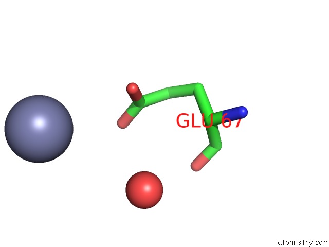
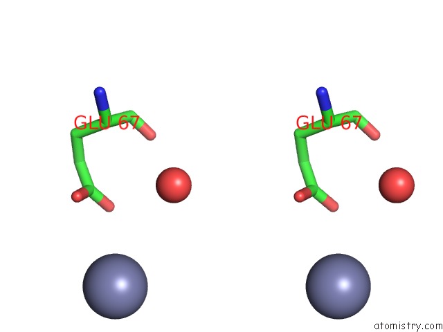
Zinc binding site 1 out of 3 in 3tn2
Go back to
Zinc binding site 1 out
of 3 in the Structure Analysis of MIP1-Beta P8A
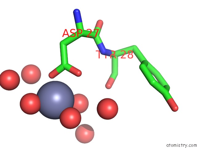
Mono view
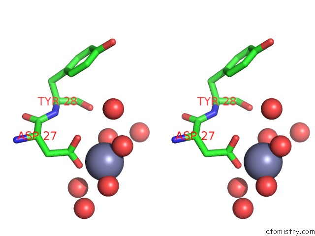
Stereo pair view
Mono view
Stereo pair view
A full contact list of Zinc with other atoms in the Zn binding
site number 1 of Structure Analysis of MIP1-Beta P8A within 5.0Å range:
|
Zinc binding site 2 out of 3 in 3tn2
Go back to
Zinc binding site 2 out
of 3 in the Structure Analysis of MIP1-Beta P8A
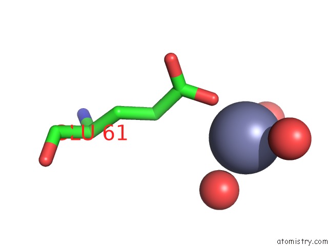
Mono view
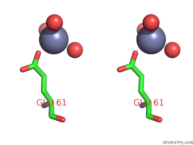
Stereo pair view
Mono view
Stereo pair view
A full contact list of Zinc with other atoms in the Zn binding
site number 2 of Structure Analysis of MIP1-Beta P8A within 5.0Å range:
|
Zinc binding site 3 out of 3 in 3tn2
Go back to
Zinc binding site 3 out
of 3 in the Structure Analysis of MIP1-Beta P8A
Mono view
Stereo pair view
Mono view
Stereo pair view
A full contact list of Zinc with other atoms in the Zn binding
site number 3 of Structure Analysis of MIP1-Beta P8A within 5.0Å range:
|
Reference:
W.G.Liang,
M.Ren,
F.Zhao,
W.J.Tang.
Structures of Human CCL18, CCL3, and CCL4 Reveal Molecular Determinants For Quaternary Structures and Sensitivity to Insulin-Degrading Enzyme. J.Mol.Biol. V. 427 1345 2015.
ISSN: ISSN 0022-2836
PubMed: 25636406
DOI: 10.1016/J.JMB.2015.01.012
Page generated: Sat Oct 26 16:38:42 2024
ISSN: ISSN 0022-2836
PubMed: 25636406
DOI: 10.1016/J.JMB.2015.01.012
Last articles
Zn in 9MJ5Zn in 9HNW
Zn in 9G0L
Zn in 9FNE
Zn in 9DZN
Zn in 9E0I
Zn in 9D32
Zn in 9DAK
Zn in 8ZXC
Zn in 8ZUF