Zinc »
PDB 3sje-3sue »
3sp4 »
Zinc in PDB 3sp4: Crystal Structure of Aprataxin Ortholog HNT3 From Schizosaccharomyces Pombe
Protein crystallography data
The structure of Crystal Structure of Aprataxin Ortholog HNT3 From Schizosaccharomyces Pombe, PDB code: 3sp4
was solved by
Y.Gong,
D.Zhu,
J.Ding,
C.Dou,
X.Ren,
T.Jiang,
D.Wang,
with X-Ray Crystallography technique. A brief refinement statistics is given in the table below:
| Resolution Low / High (Å) | 37.86 / 1.80 |
| Space group | P 21 21 21 |
| Cell size a, b, c (Å), α, β, γ (°) | 53.177, 70.201, 107.809, 90.00, 90.00, 90.00 |
| R / Rfree (%) | 18.5 / 22.5 |
Zinc Binding Sites:
The binding sites of Zinc atom in the Crystal Structure of Aprataxin Ortholog HNT3 From Schizosaccharomyces Pombe
(pdb code 3sp4). This binding sites where shown within
5.0 Angstroms radius around Zinc atom.
In total 2 binding sites of Zinc where determined in the Crystal Structure of Aprataxin Ortholog HNT3 From Schizosaccharomyces Pombe, PDB code: 3sp4:
Jump to Zinc binding site number: 1; 2;
In total 2 binding sites of Zinc where determined in the Crystal Structure of Aprataxin Ortholog HNT3 From Schizosaccharomyces Pombe, PDB code: 3sp4:
Jump to Zinc binding site number: 1; 2;
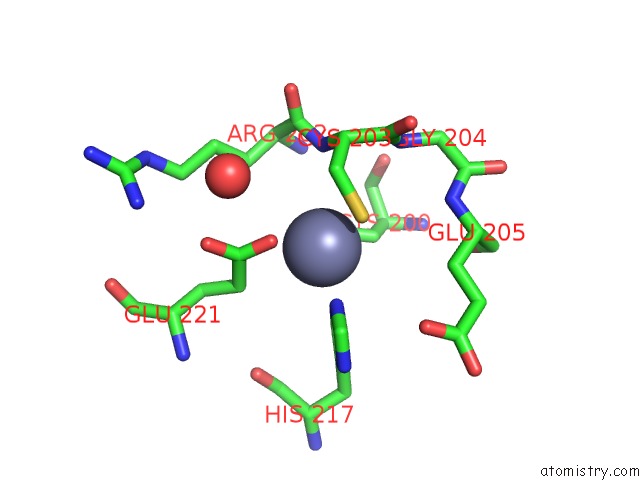
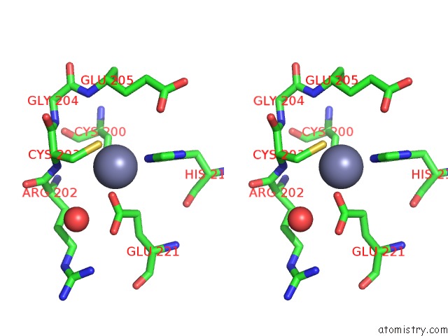
Zinc binding site 1 out of 2 in 3sp4
Go back to
Zinc binding site 1 out
of 2 in the Crystal Structure of Aprataxin Ortholog HNT3 From Schizosaccharomyces Pombe
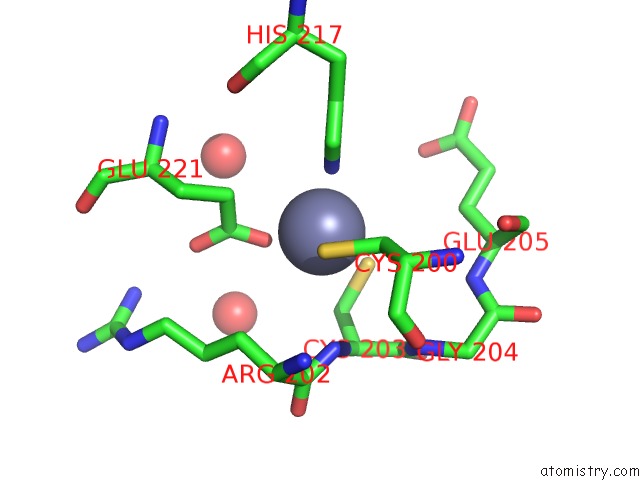
Mono view

Stereo pair view
Mono view
Stereo pair view
A full contact list of Zinc with other atoms in the Zn binding
site number 1 of Crystal Structure of Aprataxin Ortholog HNT3 From Schizosaccharomyces Pombe within 5.0Å range:
|
Zinc binding site 2 out of 2 in 3sp4
Go back to
Zinc binding site 2 out
of 2 in the Crystal Structure of Aprataxin Ortholog HNT3 From Schizosaccharomyces Pombe
Mono view
Stereo pair view
Mono view
Stereo pair view
A full contact list of Zinc with other atoms in the Zn binding
site number 2 of Crystal Structure of Aprataxin Ortholog HNT3 From Schizosaccharomyces Pombe within 5.0Å range:
|
Reference:
Y.Gong,
D.Zhu,
J.Ding,
C.Dou,
X.Ren,
L.Gu,
T.Jiang,
D.Wang.
Crystal Structures of Aprataxin Ortholog HNT3 Reveal the Mechanism For Reversal of 5'-Adenylated Dna Nat.Struct.Mol.Biol. V. 18 1297 2011.
ISSN: ISSN 1545-9993
PubMed: 21984208
DOI: 10.1038/NSMB.2145
Page generated: Sat Oct 26 16:01:45 2024
ISSN: ISSN 1545-9993
PubMed: 21984208
DOI: 10.1038/NSMB.2145
Last articles
Zn in 9MJ5Zn in 9HNW
Zn in 9G0L
Zn in 9FNE
Zn in 9DZN
Zn in 9E0I
Zn in 9D32
Zn in 9DAK
Zn in 8ZXC
Zn in 8ZUF