Zinc »
PDB 3s2s-3scj »
3s2s »
Zinc in PDB 3s2s: The Crystal Structure of Pyrazinamidase/Nicotinamidase From Streptococcus Mutans UA159
Enzymatic activity of The Crystal Structure of Pyrazinamidase/Nicotinamidase From Streptococcus Mutans UA159
All present enzymatic activity of The Crystal Structure of Pyrazinamidase/Nicotinamidase From Streptococcus Mutans UA159:
3.5.1.19;
3.5.1.19;
Protein crystallography data
The structure of The Crystal Structure of Pyrazinamidase/Nicotinamidase From Streptococcus Mutans UA159, PDB code: 3s2s
was solved by
X.-D.Su,
X.Liu,
H.Zhang,
with X-Ray Crystallography technique. A brief refinement statistics is given in the table below:
| Resolution Low / High (Å) | 19.98 / 1.70 |
| Space group | P 21 21 21 |
| Cell size a, b, c (Å), α, β, γ (°) | 76.490, 80.120, 130.960, 90.00, 90.00, 90.00 |
| R / Rfree (%) | 16 / 19.1 |
Other elements in 3s2s:
The structure of The Crystal Structure of Pyrazinamidase/Nicotinamidase From Streptococcus Mutans UA159 also contains other interesting chemical elements:
| Arsenic | (As) | 4 atoms |
Zinc Binding Sites:
The binding sites of Zinc atom in the The Crystal Structure of Pyrazinamidase/Nicotinamidase From Streptococcus Mutans UA159
(pdb code 3s2s). This binding sites where shown within
5.0 Angstroms radius around Zinc atom.
In total 4 binding sites of Zinc where determined in the The Crystal Structure of Pyrazinamidase/Nicotinamidase From Streptococcus Mutans UA159, PDB code: 3s2s:
Jump to Zinc binding site number: 1; 2; 3; 4;
In total 4 binding sites of Zinc where determined in the The Crystal Structure of Pyrazinamidase/Nicotinamidase From Streptococcus Mutans UA159, PDB code: 3s2s:
Jump to Zinc binding site number: 1; 2; 3; 4;
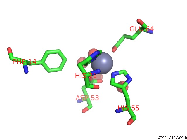
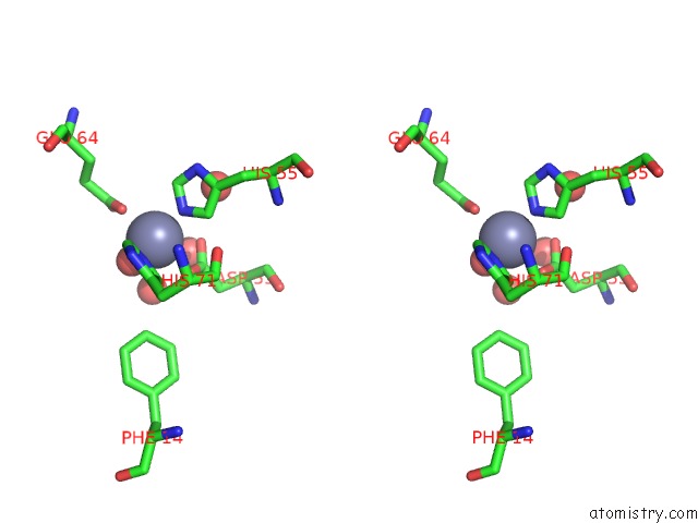
Zinc binding site 1 out of 4 in 3s2s
Go back to
Zinc binding site 1 out
of 4 in the The Crystal Structure of Pyrazinamidase/Nicotinamidase From Streptococcus Mutans UA159
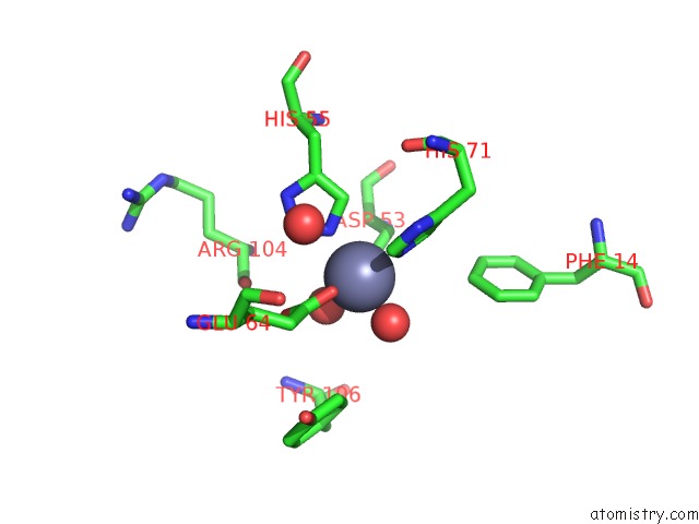
Mono view
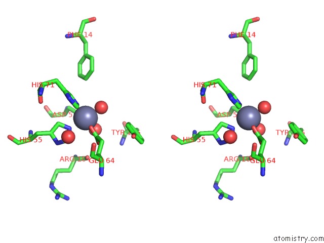
Stereo pair view
Mono view
Stereo pair view
A full contact list of Zinc with other atoms in the Zn binding
site number 1 of The Crystal Structure of Pyrazinamidase/Nicotinamidase From Streptococcus Mutans UA159 within 5.0Å range:
|
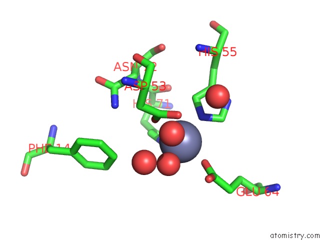
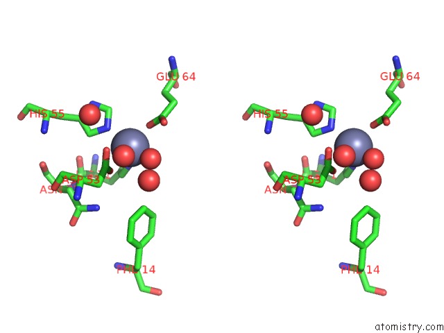
Zinc binding site 2 out of 4 in 3s2s
Go back to
Zinc binding site 2 out
of 4 in the The Crystal Structure of Pyrazinamidase/Nicotinamidase From Streptococcus Mutans UA159
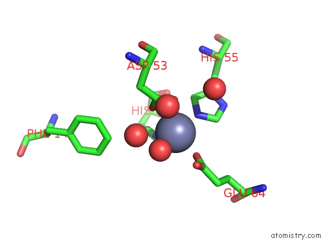
Mono view
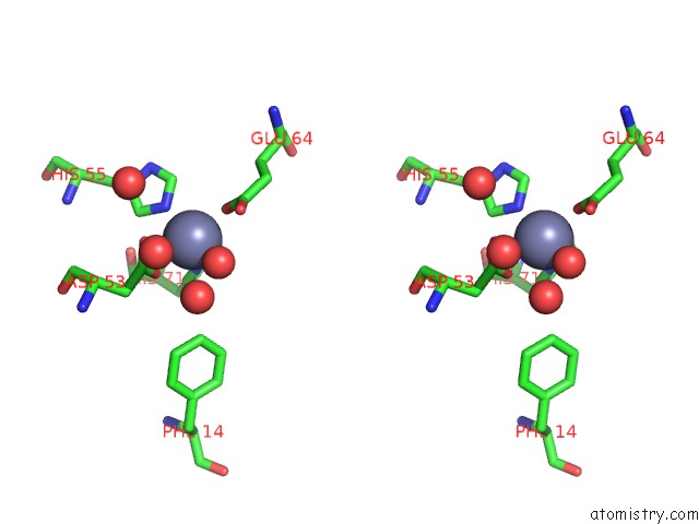
Stereo pair view
Mono view
Stereo pair view
A full contact list of Zinc with other atoms in the Zn binding
site number 2 of The Crystal Structure of Pyrazinamidase/Nicotinamidase From Streptococcus Mutans UA159 within 5.0Å range:
|
Zinc binding site 3 out of 4 in 3s2s
Go back to
Zinc binding site 3 out
of 4 in the The Crystal Structure of Pyrazinamidase/Nicotinamidase From Streptococcus Mutans UA159
Mono view
Stereo pair view
Mono view
Stereo pair view
A full contact list of Zinc with other atoms in the Zn binding
site number 3 of The Crystal Structure of Pyrazinamidase/Nicotinamidase From Streptococcus Mutans UA159 within 5.0Å range:
|
Zinc binding site 4 out of 4 in 3s2s
Go back to
Zinc binding site 4 out
of 4 in the The Crystal Structure of Pyrazinamidase/Nicotinamidase From Streptococcus Mutans UA159
Mono view
Stereo pair view
Mono view
Stereo pair view
A full contact list of Zinc with other atoms in the Zn binding
site number 4 of The Crystal Structure of Pyrazinamidase/Nicotinamidase From Streptococcus Mutans UA159 within 5.0Å range:
|
Reference:
X.Liu,
H.Zhang,
X.J.Wang,
L.F.Li,
X.-D.Su.
Get Phases From Arsenic Anomalous Scattering: De Novo Sad Phasing of Two Protein Structures Crystallized in Cacodylate Buffer Plos One V. 6 24227 2011.
ISSN: ESSN 1932-6203
PubMed: 21912678
DOI: 10.1371/JOURNAL.PONE.0024227
Page generated: Sat Oct 26 15:33:02 2024
ISSN: ESSN 1932-6203
PubMed: 21912678
DOI: 10.1371/JOURNAL.PONE.0024227
Last articles
Zn in 9MJ5Zn in 9HNW
Zn in 9G0L
Zn in 9FNE
Zn in 9DZN
Zn in 9E0I
Zn in 9D32
Zn in 9DAK
Zn in 8ZXC
Zn in 8ZUF