Zinc »
PDB 3rjw-3ryj »
3rq4 »
Zinc in PDB 3rq4: Crystal Structure of Suppressor of Variegation 4-20 Homolog 2
Enzymatic activity of Crystal Structure of Suppressor of Variegation 4-20 Homolog 2
All present enzymatic activity of Crystal Structure of Suppressor of Variegation 4-20 Homolog 2:
2.1.1.43;
2.1.1.43;
Protein crystallography data
The structure of Crystal Structure of Suppressor of Variegation 4-20 Homolog 2, PDB code: 3rq4
was solved by
A.Dong,
H.Zeng,
W.Tempel,
P.Loppnau,
C.Bountra,
J.Weigelt,
C.H.Arrowsmith,
A.M.Edwards,
J.Min,
H.Wu,
Structural Genomics Consortium (Sgc),
with X-Ray Crystallography technique. A brief refinement statistics is given in the table below:
| Resolution Low / High (Å) | 50.00 / 1.80 |
| Space group | P 21 21 21 |
| Cell size a, b, c (Å), α, β, γ (°) | 34.680, 60.151, 126.601, 90.00, 90.00, 90.00 |
| R / Rfree (%) | 17.8 / 22.1 |
Zinc Binding Sites:
The binding sites of Zinc atom in the Crystal Structure of Suppressor of Variegation 4-20 Homolog 2
(pdb code 3rq4). This binding sites where shown within
5.0 Angstroms radius around Zinc atom.
In total only one binding site of Zinc was determined in the Crystal Structure of Suppressor of Variegation 4-20 Homolog 2, PDB code: 3rq4:
In total only one binding site of Zinc was determined in the Crystal Structure of Suppressor of Variegation 4-20 Homolog 2, PDB code: 3rq4:
Zinc binding site 1 out of 1 in 3rq4
Go back to
Zinc binding site 1 out
of 1 in the Crystal Structure of Suppressor of Variegation 4-20 Homolog 2
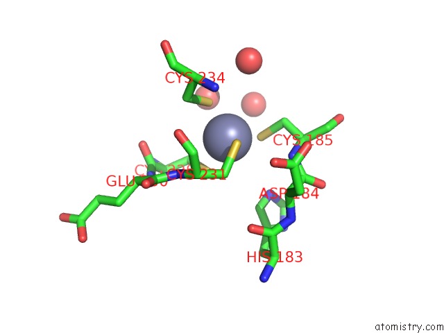
Mono view
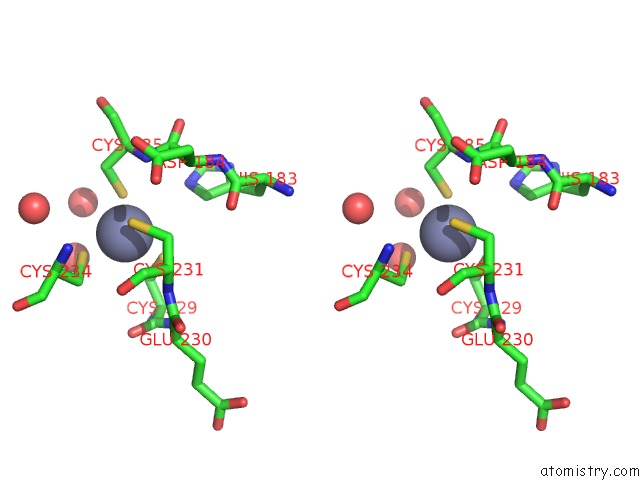
Stereo pair view
Mono view
Stereo pair view
A full contact list of Zinc with other atoms in the Zn binding
site number 1 of Crystal Structure of Suppressor of Variegation 4-20 Homolog 2 within 5.0Å range:
|
Reference:
H.Wu,
A.Siarheyeva,
H.Zeng,
R.Lam,
A.Dong,
X.H.Wu,
Y.Li,
M.Schapira,
M.Vedadi,
J.Min.
Crystal Structures of the Human Histone H4K20 Methyltransferases SUV420H1 and SUV420H2. Febs Lett. V. 587 3859 2013.
ISSN: ISSN 0014-5793
PubMed: 24396869
Page generated: Wed Aug 20 13:38:12 2025
ISSN: ISSN 0014-5793
PubMed: 24396869
Last articles
Zn in 4K60Zn in 4K5P
Zn in 4K5Z
Zn in 4K5O
Zn in 4K5N
Zn in 4K5M
Zn in 4K5L
Zn in 4K5I
Zn in 4K5K
Zn in 4K5J