Zinc »
PDB 3qmc-3qzv »
3qna »
Zinc in PDB 3qna: Crystal Structure of A 6-Pyruvoyltetrahydropterin Synthase Homologue From Esherichia Coli Complexed Sepiapterin
Enzymatic activity of Crystal Structure of A 6-Pyruvoyltetrahydropterin Synthase Homologue From Esherichia Coli Complexed Sepiapterin
All present enzymatic activity of Crystal Structure of A 6-Pyruvoyltetrahydropterin Synthase Homologue From Esherichia Coli Complexed Sepiapterin:
4.1.2.50;
4.1.2.50;
Protein crystallography data
The structure of Crystal Structure of A 6-Pyruvoyltetrahydropterin Synthase Homologue From Esherichia Coli Complexed Sepiapterin, PDB code: 3qna
was solved by
K.H.Seo,
N.N.Zhuang,
K.H.Lee,
with X-Ray Crystallography technique. A brief refinement statistics is given in the table below:
| Resolution Low / High (Å) | 92.85 / 2.50 |
| Space group | I 2 2 2 |
| Cell size a, b, c (Å), α, β, γ (°) | 112.714, 115.882, 155.185, 90.00, 90.00, 90.00 |
| R / Rfree (%) | 18.2 / 23 |
Zinc Binding Sites:
The binding sites of Zinc atom in the Crystal Structure of A 6-Pyruvoyltetrahydropterin Synthase Homologue From Esherichia Coli Complexed Sepiapterin
(pdb code 3qna). This binding sites where shown within
5.0 Angstroms radius around Zinc atom.
In total 6 binding sites of Zinc where determined in the Crystal Structure of A 6-Pyruvoyltetrahydropterin Synthase Homologue From Esherichia Coli Complexed Sepiapterin, PDB code: 3qna:
Jump to Zinc binding site number: 1; 2; 3; 4; 5; 6;
In total 6 binding sites of Zinc where determined in the Crystal Structure of A 6-Pyruvoyltetrahydropterin Synthase Homologue From Esherichia Coli Complexed Sepiapterin, PDB code: 3qna:
Jump to Zinc binding site number: 1; 2; 3; 4; 5; 6;
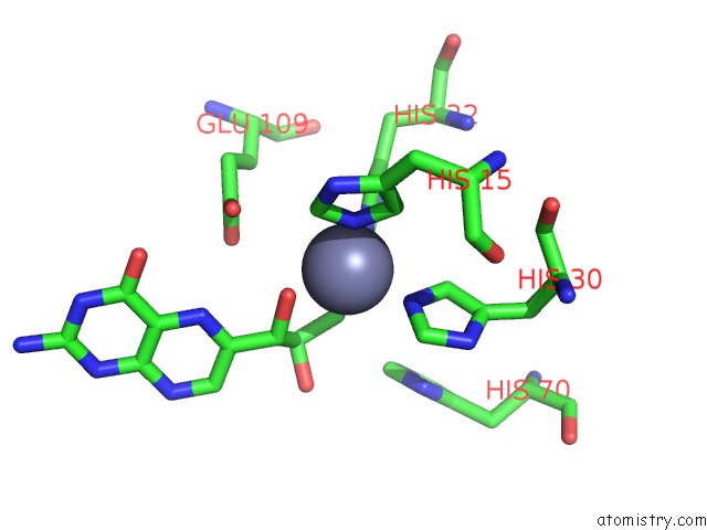
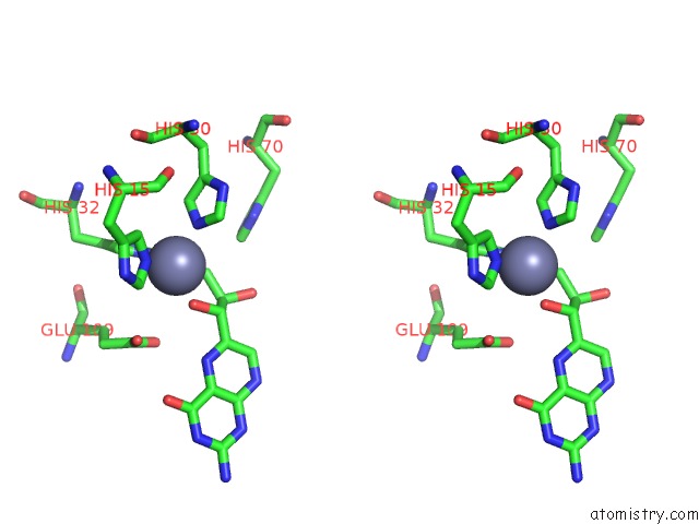
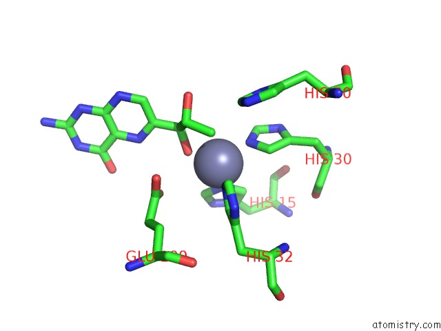
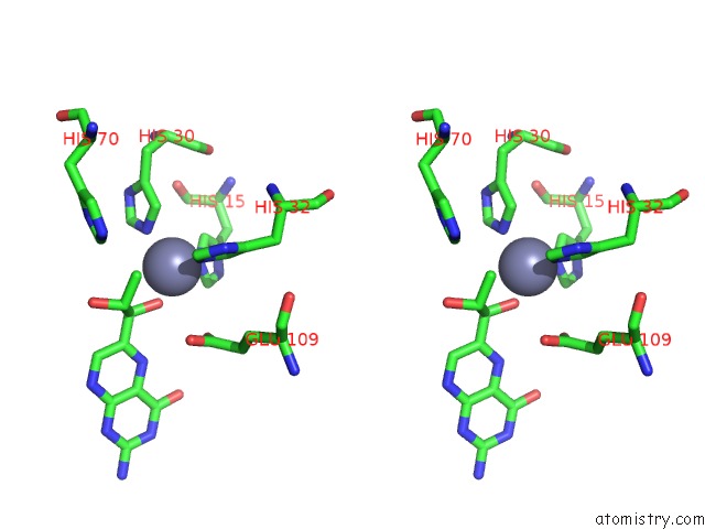
Zinc binding site 1 out of 6 in 3qna
Go back to
Zinc binding site 1 out
of 6 in the Crystal Structure of A 6-Pyruvoyltetrahydropterin Synthase Homologue From Esherichia Coli Complexed Sepiapterin
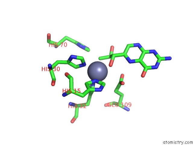
Mono view
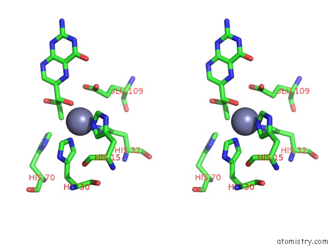
Stereo pair view
Mono view
Stereo pair view
A full contact list of Zinc with other atoms in the Zn binding
site number 1 of Crystal Structure of A 6-Pyruvoyltetrahydropterin Synthase Homologue From Esherichia Coli Complexed Sepiapterin within 5.0Å range:
|
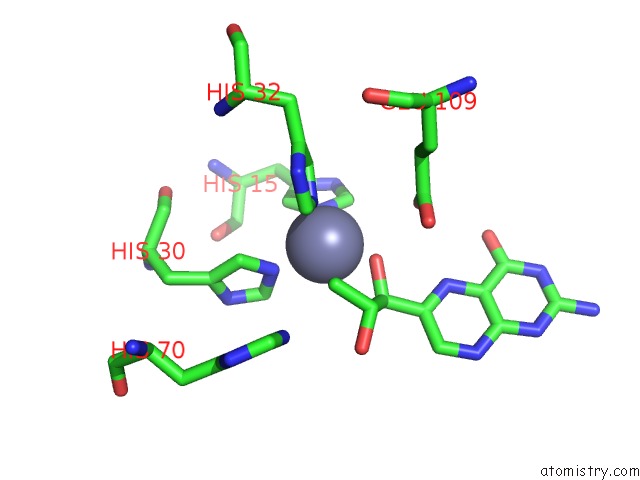
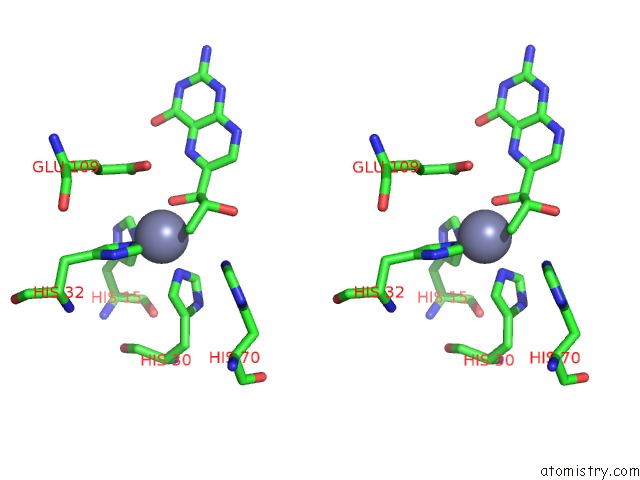
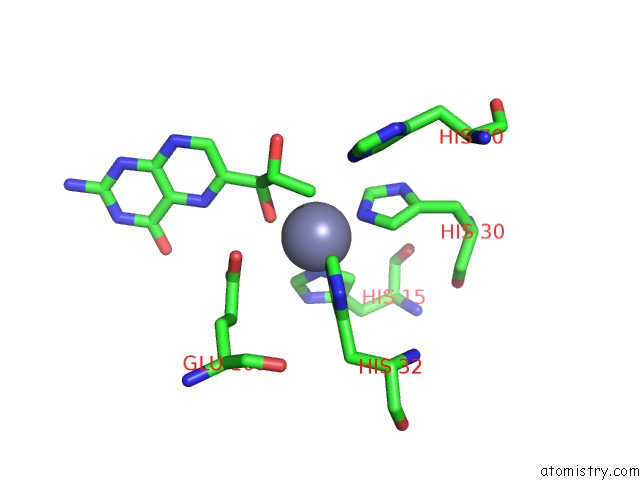
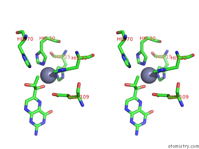
Zinc binding site 2 out of 6 in 3qna
Go back to
Zinc binding site 2 out
of 6 in the Crystal Structure of A 6-Pyruvoyltetrahydropterin Synthase Homologue From Esherichia Coli Complexed Sepiapterin
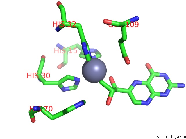
Mono view
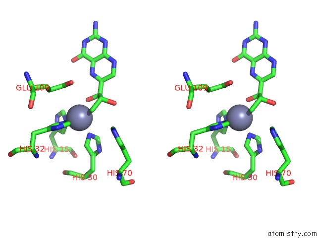
Stereo pair view
Mono view
Stereo pair view
A full contact list of Zinc with other atoms in the Zn binding
site number 2 of Crystal Structure of A 6-Pyruvoyltetrahydropterin Synthase Homologue From Esherichia Coli Complexed Sepiapterin within 5.0Å range:
|
Zinc binding site 3 out of 6 in 3qna
Go back to
Zinc binding site 3 out
of 6 in the Crystal Structure of A 6-Pyruvoyltetrahydropterin Synthase Homologue From Esherichia Coli Complexed Sepiapterin
Mono view
Stereo pair view
Mono view
Stereo pair view
A full contact list of Zinc with other atoms in the Zn binding
site number 3 of Crystal Structure of A 6-Pyruvoyltetrahydropterin Synthase Homologue From Esherichia Coli Complexed Sepiapterin within 5.0Å range:
|
Zinc binding site 4 out of 6 in 3qna
Go back to
Zinc binding site 4 out
of 6 in the Crystal Structure of A 6-Pyruvoyltetrahydropterin Synthase Homologue From Esherichia Coli Complexed Sepiapterin
Mono view
Stereo pair view
Mono view
Stereo pair view
A full contact list of Zinc with other atoms in the Zn binding
site number 4 of Crystal Structure of A 6-Pyruvoyltetrahydropterin Synthase Homologue From Esherichia Coli Complexed Sepiapterin within 5.0Å range:
|
Zinc binding site 5 out of 6 in 3qna
Go back to
Zinc binding site 5 out
of 6 in the Crystal Structure of A 6-Pyruvoyltetrahydropterin Synthase Homologue From Esherichia Coli Complexed Sepiapterin
Mono view
Stereo pair view
Mono view
Stereo pair view
A full contact list of Zinc with other atoms in the Zn binding
site number 5 of Crystal Structure of A 6-Pyruvoyltetrahydropterin Synthase Homologue From Esherichia Coli Complexed Sepiapterin within 5.0Å range:
|
Zinc binding site 6 out of 6 in 3qna
Go back to
Zinc binding site 6 out
of 6 in the Crystal Structure of A 6-Pyruvoyltetrahydropterin Synthase Homologue From Esherichia Coli Complexed Sepiapterin
Mono view
Stereo pair view
Mono view
Stereo pair view
A full contact list of Zinc with other atoms in the Zn binding
site number 6 of Crystal Structure of A 6-Pyruvoyltetrahydropterin Synthase Homologue From Esherichia Coli Complexed Sepiapterin within 5.0Å range:
|
Reference:
K.H.Seo,
N.Zhuang,
Y.S.Park,
K.H.Park,
K.H.Lee.
Structural Basis of A Novel Activity of Bacterial 6-Pyruvoyltetrahydropterin Synthase Homologues Distinct From Mammalian 6-Pyruvoyltetrahydropterin Synthase Activity. Acta Crystallogr.,Sect.D V. 70 1212 2014.
ISSN: ISSN 0907-4449
PubMed: 24816091
DOI: 10.1107/S1399004714002016
Page generated: Sat Oct 26 12:14:34 2024
ISSN: ISSN 0907-4449
PubMed: 24816091
DOI: 10.1107/S1399004714002016
Last articles
Zn in 9MJ5Zn in 9HNW
Zn in 9G0L
Zn in 9FNE
Zn in 9DZN
Zn in 9E0I
Zn in 9D32
Zn in 9DAK
Zn in 8ZXC
Zn in 8ZUF