Zinc »
PDB 3q87-3qmb »
3qiz »
Zinc in PDB 3qiz: Crystal Structure of Bont/A Lc Complexed with Hydroxamate-Based Inhibitor Pt-2
Enzymatic activity of Crystal Structure of Bont/A Lc Complexed with Hydroxamate-Based Inhibitor Pt-2
All present enzymatic activity of Crystal Structure of Bont/A Lc Complexed with Hydroxamate-Based Inhibitor Pt-2:
3.4.24.69;
3.4.24.69;
Protein crystallography data
The structure of Crystal Structure of Bont/A Lc Complexed with Hydroxamate-Based Inhibitor Pt-2, PDB code: 3qiz
was solved by
A.A.Thompson,
G.W.Han,
R.C.Stevens,
with X-Ray Crystallography technique. A brief refinement statistics is given in the table below:
| Resolution Low / High (Å) | 37.13 / 2.00 |
| Space group | P 21 21 2 |
| Cell size a, b, c (Å), α, β, γ (°) | 59.230, 190.654, 42.430, 90.00, 90.00, 90.00 |
| R / Rfree (%) | 18.8 / 22.3 |
Other elements in 3qiz:
The structure of Crystal Structure of Bont/A Lc Complexed with Hydroxamate-Based Inhibitor Pt-2 also contains other interesting chemical elements:
| Fluorine | (F) | 2 atoms |
Zinc Binding Sites:
The binding sites of Zinc atom in the Crystal Structure of Bont/A Lc Complexed with Hydroxamate-Based Inhibitor Pt-2
(pdb code 3qiz). This binding sites where shown within
5.0 Angstroms radius around Zinc atom.
In total only one binding site of Zinc was determined in the Crystal Structure of Bont/A Lc Complexed with Hydroxamate-Based Inhibitor Pt-2, PDB code: 3qiz:
In total only one binding site of Zinc was determined in the Crystal Structure of Bont/A Lc Complexed with Hydroxamate-Based Inhibitor Pt-2, PDB code: 3qiz:
Zinc binding site 1 out of 1 in 3qiz
Go back to
Zinc binding site 1 out
of 1 in the Crystal Structure of Bont/A Lc Complexed with Hydroxamate-Based Inhibitor Pt-2
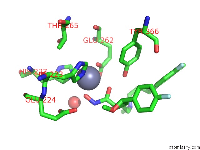
Mono view
Stereo pair view
Mono view
Stereo pair view
A full contact list of Zinc with other atoms in the Zn binding
site number 1 of Crystal Structure of Bont/A Lc Complexed with Hydroxamate-Based Inhibitor Pt-2 within 5.0Å range:
|
Reference:
A.A.Thompson,
G.S.Jiao,
S.Kim,
A.Thai,
L.Cregar-Hernandez,
S.A.Margosiak,
A.T.Johnson,
G.W.Han,
S.O'malley,
R.C.Stevens.
Structural Characterization of Three Novel Hydroxamate-Based Zinc Chelating Inhibitors of the Clostridium Botulinum Serotype A Neurotoxin Light Chain Metalloprotease Reveals A Compact Binding Site Resulting From 60/70 Loop Flexibility. Biochemistry V. 50 4019 2011.
ISSN: ISSN 0006-2960
PubMed: 21434688
DOI: 10.1021/BI2001483
Page generated: Sat Oct 26 12:07:53 2024
ISSN: ISSN 0006-2960
PubMed: 21434688
DOI: 10.1021/BI2001483
Last articles
Zn in 9MJ5Zn in 9HNW
Zn in 9G0L
Zn in 9FNE
Zn in 9DZN
Zn in 9E0I
Zn in 9D32
Zn in 9DAK
Zn in 8ZXC
Zn in 8ZUF