Zinc »
PDB 3phx-3ptm »
3psr »
Zinc in PDB 3psr: Human Psoriasin (S100A7) CA2+ Bound Form (Crystal Form I)
Protein crystallography data
The structure of Human Psoriasin (S100A7) CA2+ Bound Form (Crystal Form I), PDB code: 3psr
was solved by
D.E.Brodersen,
J.Nyborg,
M.Kjeldgaard,
with X-Ray Crystallography technique. A brief refinement statistics is given in the table below:
| Resolution Low / High (Å) | 100.00 / 2.50 |
| Space group | P 21 21 21 |
| Cell size a, b, c (Å), α, β, γ (°) | 52.150, 56.670, 76.380, 90.00, 90.00, 90.00 |
| R / Rfree (%) | 22 / 29.5 |
Other elements in 3psr:
The structure of Human Psoriasin (S100A7) CA2+ Bound Form (Crystal Form I) also contains other interesting chemical elements:
| Calcium | (Ca) | 2 atoms |
Zinc Binding Sites:
The binding sites of Zinc atom in the Human Psoriasin (S100A7) CA2+ Bound Form (Crystal Form I)
(pdb code 3psr). This binding sites where shown within
5.0 Angstroms radius around Zinc atom.
In total only one binding site of Zinc was determined in the Human Psoriasin (S100A7) CA2+ Bound Form (Crystal Form I), PDB code: 3psr:
In total only one binding site of Zinc was determined in the Human Psoriasin (S100A7) CA2+ Bound Form (Crystal Form I), PDB code: 3psr:
Zinc binding site 1 out of 1 in 3psr
Go back to
Zinc binding site 1 out
of 1 in the Human Psoriasin (S100A7) CA2+ Bound Form (Crystal Form I)
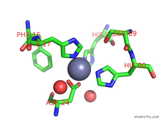
Mono view
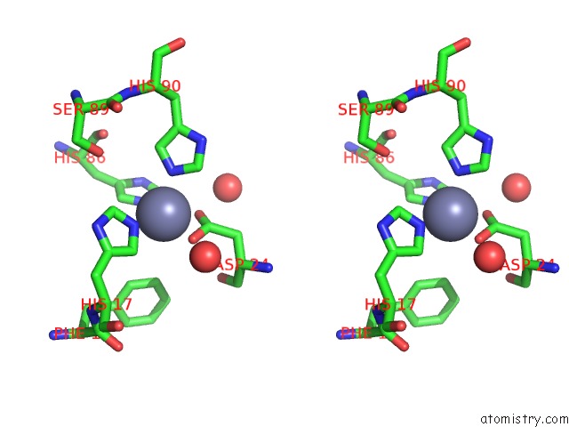
Stereo pair view
Mono view
Stereo pair view
A full contact list of Zinc with other atoms in the Zn binding
site number 1 of Human Psoriasin (S100A7) CA2+ Bound Form (Crystal Form I) within 5.0Å range:
|
Reference:
D.E.Brodersen,
J.Nyborg,
M.Kjeldgaard.
Zinc-Binding Site of An S100 Protein Revealed. Two Crystal Structures of CA2+-Bound Human Psoriasin (S100A7) in the ZN2+-Loaded and ZN2+-Free States. Biochemistry V. 38 1695 1999.
ISSN: ISSN 0006-2960
PubMed: 10026247
DOI: 10.1021/BI982483D
Page generated: Sat Oct 26 11:41:21 2024
ISSN: ISSN 0006-2960
PubMed: 10026247
DOI: 10.1021/BI982483D
Last articles
Zn in 9MJ5Zn in 9HNW
Zn in 9G0L
Zn in 9FNE
Zn in 9DZN
Zn in 9E0I
Zn in 9D32
Zn in 9DAK
Zn in 8ZXC
Zn in 8ZUF