Zinc »
PDB 3phx-3ptm »
3ps1 »
Zinc in PDB 3ps1: Crystal Structure of the Escherichia Coli Lpxc/Lpc-011 Complex
Protein crystallography data
The structure of Crystal Structure of the Escherichia Coli Lpxc/Lpc-011 Complex, PDB code: 3ps1
was solved by
C.-J.Lee,
P.Zhou,
with X-Ray Crystallography technique. A brief refinement statistics is given in the table below:
| Resolution Low / High (Å) | 23.19 / 1.85 |
| Space group | P 61 |
| Cell size a, b, c (Å), α, β, γ (°) | 107.130, 107.130, 52.087, 90.00, 90.00, 120.00 |
| R / Rfree (%) | 19.3 / 21 |
Zinc Binding Sites:
The binding sites of Zinc atom in the Crystal Structure of the Escherichia Coli Lpxc/Lpc-011 Complex
(pdb code 3ps1). This binding sites where shown within
5.0 Angstroms radius around Zinc atom.
In total only one binding site of Zinc was determined in the Crystal Structure of the Escherichia Coli Lpxc/Lpc-011 Complex, PDB code: 3ps1:
In total only one binding site of Zinc was determined in the Crystal Structure of the Escherichia Coli Lpxc/Lpc-011 Complex, PDB code: 3ps1:
Zinc binding site 1 out of 1 in 3ps1
Go back to
Zinc binding site 1 out
of 1 in the Crystal Structure of the Escherichia Coli Lpxc/Lpc-011 Complex
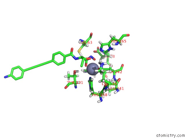
Mono view
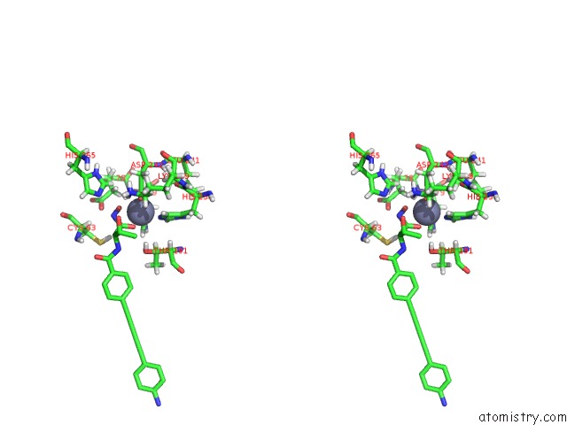
Stereo pair view
Mono view
Stereo pair view
A full contact list of Zinc with other atoms in the Zn binding
site number 1 of Crystal Structure of the Escherichia Coli Lpxc/Lpc-011 Complex within 5.0Å range:
|
Reference:
X.Liang,
C.J.Lee,
X.Chen,
H.S.Chung,
D.Zeng,
C.R.Raetz,
Y.Li,
P.Zhou,
E.J.Toone.
Syntheses, Structures and Antibiotic Activities of Lpxc Inhibitors Based on the Diacetylene Scaffold. Bioorg.Med.Chem. V. 19 852 2011.
ISSN: ISSN 0968-0896
PubMed: 21194954
DOI: 10.1016/J.BMC.2010.12.017
Page generated: Sat Oct 26 11:40:31 2024
ISSN: ISSN 0968-0896
PubMed: 21194954
DOI: 10.1016/J.BMC.2010.12.017
Last articles
Zn in 9MJ5Zn in 9HNW
Zn in 9G0L
Zn in 9FNE
Zn in 9DZN
Zn in 9E0I
Zn in 9D32
Zn in 9DAK
Zn in 8ZXC
Zn in 8ZUF