Zinc »
PDB 3p55-3phx »
3pdn »
Zinc in PDB 3pdn: Crystal Structure of SMYD3 in Complex with Methyltransferase Inhibitor Sinefungin
Enzymatic activity of Crystal Structure of SMYD3 in Complex with Methyltransferase Inhibitor Sinefungin
All present enzymatic activity of Crystal Structure of SMYD3 in Complex with Methyltransferase Inhibitor Sinefungin:
2.1.1.43;
2.1.1.43;
Protein crystallography data
The structure of Crystal Structure of SMYD3 in Complex with Methyltransferase Inhibitor Sinefungin, PDB code: 3pdn
was solved by
N.Sirinupong,
J.Brunzelle,
Z.Yang,
with X-Ray Crystallography technique. A brief refinement statistics is given in the table below:
| Resolution Low / High (Å) | 29.45 / 1.70 |
| Space group | P 21 21 21 |
| Cell size a, b, c (Å), α, β, γ (°) | 61.240, 66.246, 107.384, 90.00, 90.00, 90.00 |
| R / Rfree (%) | 15.5 / 18.9 |
Other elements in 3pdn:
The structure of Crystal Structure of SMYD3 in Complex with Methyltransferase Inhibitor Sinefungin also contains other interesting chemical elements:
| Magnesium | (Mg) | 2 atoms |
Zinc Binding Sites:
The binding sites of Zinc atom in the Crystal Structure of SMYD3 in Complex with Methyltransferase Inhibitor Sinefungin
(pdb code 3pdn). This binding sites where shown within
5.0 Angstroms radius around Zinc atom.
In total 3 binding sites of Zinc where determined in the Crystal Structure of SMYD3 in Complex with Methyltransferase Inhibitor Sinefungin, PDB code: 3pdn:
Jump to Zinc binding site number: 1; 2; 3;
In total 3 binding sites of Zinc where determined in the Crystal Structure of SMYD3 in Complex with Methyltransferase Inhibitor Sinefungin, PDB code: 3pdn:
Jump to Zinc binding site number: 1; 2; 3;
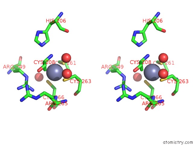
Zinc binding site 1 out of 3 in 3pdn
Go back to
Zinc binding site 1 out
of 3 in the Crystal Structure of SMYD3 in Complex with Methyltransferase Inhibitor Sinefungin

Mono view

Stereo pair view
Mono view
Stereo pair view
A full contact list of Zinc with other atoms in the Zn binding
site number 1 of Crystal Structure of SMYD3 in Complex with Methyltransferase Inhibitor Sinefungin within 5.0Å range:
|
Zinc binding site 2 out of 3 in 3pdn
Go back to
Zinc binding site 2 out
of 3 in the Crystal Structure of SMYD3 in Complex with Methyltransferase Inhibitor Sinefungin

Mono view

Stereo pair view
Mono view
Stereo pair view
A full contact list of Zinc with other atoms in the Zn binding
site number 2 of Crystal Structure of SMYD3 in Complex with Methyltransferase Inhibitor Sinefungin within 5.0Å range:
|
Zinc binding site 3 out of 3 in 3pdn
Go back to
Zinc binding site 3 out
of 3 in the Crystal Structure of SMYD3 in Complex with Methyltransferase Inhibitor Sinefungin

Mono view
Stereo pair view
Mono view
Stereo pair view
A full contact list of Zinc with other atoms in the Zn binding
site number 3 of Crystal Structure of SMYD3 in Complex with Methyltransferase Inhibitor Sinefungin within 5.0Å range:
|
Reference:
N.Sirinupong,
J.Brunzelle,
E.Doko,
Z.Yang.
Structural Insights Into the Autoinhibition and Posttranslational Activation of Histone Methyltransferase SMYD3. J.Mol.Biol. V. 406 149 2011.
ISSN: ISSN 0022-2836
PubMed: 21167177
DOI: 10.1016/J.JMB.2010.12.014
Page generated: Wed Aug 20 12:50:48 2025
ISSN: ISSN 0022-2836
PubMed: 21167177
DOI: 10.1016/J.JMB.2010.12.014
Last articles
Zn in 4IS9Zn in 4IOU
Zn in 4IR0
Zn in 4IPP
Zn in 4IN9
Zn in 4IOF
Zn in 4INS
Zn in 4IMX
Zn in 4IMU
Zn in 4IMS