Zinc »
PDB 3p4v-3phd »
3pbe »
Zinc in PDB 3pbe: Crystal Structure of the Mutant W207F of Human Secretory Glutaminyl Cyclase
Enzymatic activity of Crystal Structure of the Mutant W207F of Human Secretory Glutaminyl Cyclase
All present enzymatic activity of Crystal Structure of the Mutant W207F of Human Secretory Glutaminyl Cyclase:
2.3.2.5;
2.3.2.5;
Protein crystallography data
The structure of Crystal Structure of the Mutant W207F of Human Secretory Glutaminyl Cyclase, PDB code: 3pbe
was solved by
K.F.Huang,
S.S.Liaw,
W.L.Huang,
C.Y.Chia,
Y.C.Lo,
Y.L.Chen,
A.H.J.Wang,
with X-Ray Crystallography technique. A brief refinement statistics is given in the table below:
| Resolution Low / High (Å) | 30.00 / 1.95 |
| Space group | H 3 2 |
| Cell size a, b, c (Å), α, β, γ (°) | 118.754, 118.754, 332.093, 90.00, 90.00, 120.00 |
| R / Rfree (%) | 14.3 / 19 |
Zinc Binding Sites:
The binding sites of Zinc atom in the Crystal Structure of the Mutant W207F of Human Secretory Glutaminyl Cyclase
(pdb code 3pbe). This binding sites where shown within
5.0 Angstroms radius around Zinc atom.
In total 2 binding sites of Zinc where determined in the Crystal Structure of the Mutant W207F of Human Secretory Glutaminyl Cyclase, PDB code: 3pbe:
Jump to Zinc binding site number: 1; 2;
In total 2 binding sites of Zinc where determined in the Crystal Structure of the Mutant W207F of Human Secretory Glutaminyl Cyclase, PDB code: 3pbe:
Jump to Zinc binding site number: 1; 2;
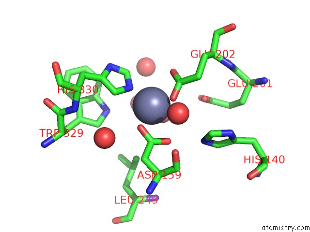
Zinc binding site 1 out of 2 in 3pbe
Go back to
Zinc binding site 1 out
of 2 in the Crystal Structure of the Mutant W207F of Human Secretory Glutaminyl Cyclase
Mono view

Stereo pair view
Mono view
Stereo pair view
A full contact list of Zinc with other atoms in the Zn binding
site number 1 of Crystal Structure of the Mutant W207F of Human Secretory Glutaminyl Cyclase within 5.0Å range:
|
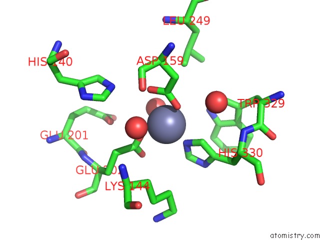
Zinc binding site 2 out of 2 in 3pbe
Go back to
Zinc binding site 2 out
of 2 in the Crystal Structure of the Mutant W207F of Human Secretory Glutaminyl Cyclase
Mono view

Stereo pair view
Mono view
Stereo pair view
A full contact list of Zinc with other atoms in the Zn binding
site number 2 of Crystal Structure of the Mutant W207F of Human Secretory Glutaminyl Cyclase within 5.0Å range:
|
Reference:
K.F.Huang,
S.S.Liaw,
W.L.Huang,
C.Y.Chia,
Y.C.Lo,
Y.L.Chen,
A.H.J.Wang.
Structures of Human Golgi-Resident Glutaminyl Cyclase and Its Complexes with Inhibitors Reveal A Large Loop Movement Upon Inhibitor Binding J.Biol.Chem. V. 286 12439 2011.
ISSN: ISSN 0021-9258
PubMed: 21288892
DOI: 10.1074/JBC.M110.208595
Page generated: Sat Oct 26 11:23:11 2024
ISSN: ISSN 0021-9258
PubMed: 21288892
DOI: 10.1074/JBC.M110.208595
Last articles
Zn in 9MJ5Zn in 9HNW
Zn in 9G0L
Zn in 9FNE
Zn in 9DZN
Zn in 9E0I
Zn in 9D32
Zn in 9DAK
Zn in 8ZXC
Zn in 8ZUF