Zinc »
PDB 3no5-3o7a »
3nos »
Zinc in PDB 3nos: Human Endothelial Nitric Oxide Synthase with Arginine Substrate
Enzymatic activity of Human Endothelial Nitric Oxide Synthase with Arginine Substrate
All present enzymatic activity of Human Endothelial Nitric Oxide Synthase with Arginine Substrate:
1.14.13.39;
1.14.13.39;
Protein crystallography data
The structure of Human Endothelial Nitric Oxide Synthase with Arginine Substrate, PDB code: 3nos
was solved by
T.O.Fischmann,
P.C.Weber,
with X-Ray Crystallography technique. A brief refinement statistics is given in the table below:
| Resolution Low / High (Å) | 8.00 / 2.40 |
| Space group | P 21 21 21 |
| Cell size a, b, c (Å), α, β, γ (°) | 68.859, 93.264, 156.117, 90.00, 90.00, 90.00 |
| R / Rfree (%) | 19.3 / 30.8 |
Other elements in 3nos:
The structure of Human Endothelial Nitric Oxide Synthase with Arginine Substrate also contains other interesting chemical elements:
| Iron | (Fe) | 2 atoms |
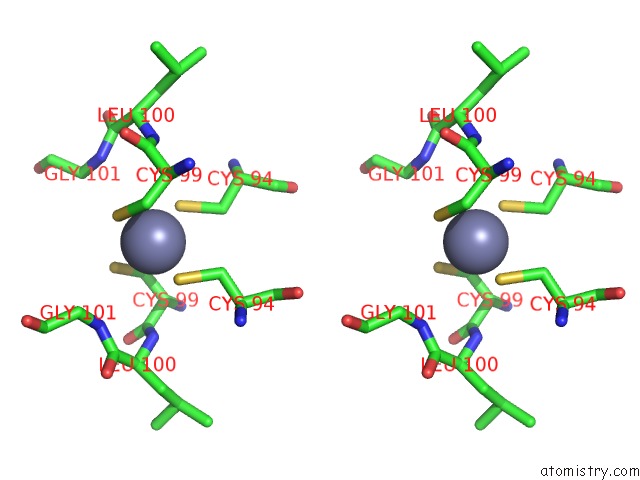
Zinc Binding Sites:
The binding sites of Zinc atom in the Human Endothelial Nitric Oxide Synthase with Arginine Substrate
(pdb code 3nos). This binding sites where shown within
5.0 Angstroms radius around Zinc atom.
In total only one binding site of Zinc was determined in the Human Endothelial Nitric Oxide Synthase with Arginine Substrate, PDB code: 3nos:
In total only one binding site of Zinc was determined in the Human Endothelial Nitric Oxide Synthase with Arginine Substrate, PDB code: 3nos:
Zinc binding site 1 out of 1 in 3nos
Go back to
Zinc binding site 1 out
of 1 in the Human Endothelial Nitric Oxide Synthase with Arginine Substrate
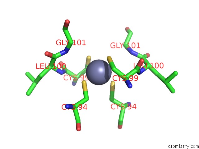
Mono view
Stereo pair view
Mono view
Stereo pair view
A full contact list of Zinc with other atoms in the Zn binding
site number 1 of Human Endothelial Nitric Oxide Synthase with Arginine Substrate within 5.0Å range:
|
Reference:
T.O.Fischmann,
A.Hruza,
X.D.Niu,
J.D.Fossetta,
C.A.Lunn,
E.Dolphin,
A.J.Prongay,
P.Reichert,
D.J.Lundell,
S.K.Narula,
P.C.Weber.
Structural Characterization of Nitric Oxide Synthase Isoforms Reveals Striking Active-Site Conservation. Nat.Struct.Biol. V. 6 233 1999.
ISSN: ISSN 1072-8368
PubMed: 10074942
DOI: 10.1038/6675
Page generated: Wed Aug 20 12:23:10 2025
ISSN: ISSN 1072-8368
PubMed: 10074942
DOI: 10.1038/6675
Last articles
Zn in 4I2HZn in 4I2F
Zn in 4I2D
Zn in 4I28
Zn in 4I1S
Zn in 4I1C
Zn in 4I15
Zn in 4I14
Zn in 4I11
Zn in 4I12