Zinc »
PDB 3m1n-3m6q »
3m3k »
Zinc in PDB 3m3k: Ligand Binding Domain (S1S2) of GLUA3 (Flop)
Protein crystallography data
The structure of Ligand Binding Domain (S1S2) of GLUA3 (Flop), PDB code: 3m3k
was solved by
A.H.Ahmed,
C.P.Ptak,
R.E.Oswald,
with X-Ray Crystallography technique. A brief refinement statistics is given in the table below:
| Resolution Low / High (Å) | 28.73 / 1.79 |
| Space group | P 2 21 21 |
| Cell size a, b, c (Å), α, β, γ (°) | 46.028, 110.329, 161.192, 90.00, 90.00, 90.00 |
| R / Rfree (%) | 20.1 / 24 |
Zinc Binding Sites:
The binding sites of Zinc atom in the Ligand Binding Domain (S1S2) of GLUA3 (Flop)
(pdb code 3m3k). This binding sites where shown within
5.0 Angstroms radius around Zinc atom.
In total 4 binding sites of Zinc where determined in the Ligand Binding Domain (S1S2) of GLUA3 (Flop), PDB code: 3m3k:
Jump to Zinc binding site number: 1; 2; 3; 4;
In total 4 binding sites of Zinc where determined in the Ligand Binding Domain (S1S2) of GLUA3 (Flop), PDB code: 3m3k:
Jump to Zinc binding site number: 1; 2; 3; 4;
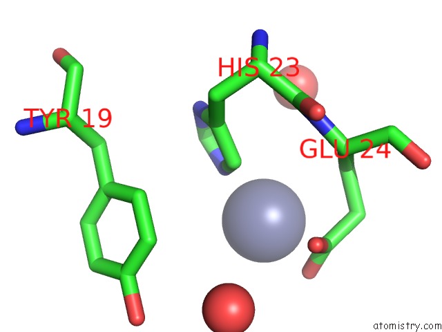
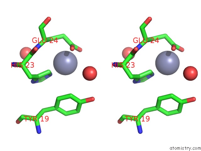
Zinc binding site 1 out of 4 in 3m3k
Go back to
Zinc binding site 1 out
of 4 in the Ligand Binding Domain (S1S2) of GLUA3 (Flop)
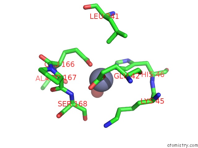
Mono view
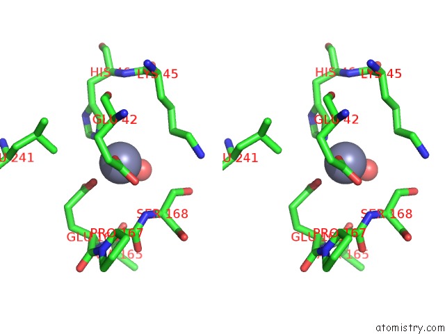
Stereo pair view
Mono view
Stereo pair view
A full contact list of Zinc with other atoms in the Zn binding
site number 1 of Ligand Binding Domain (S1S2) of GLUA3 (Flop) within 5.0Å range:
|
Zinc binding site 2 out of 4 in 3m3k
Go back to
Zinc binding site 2 out
of 4 in the Ligand Binding Domain (S1S2) of GLUA3 (Flop)
Mono view
Stereo pair view
Mono view
Stereo pair view
A full contact list of Zinc with other atoms in the Zn binding
site number 2 of Ligand Binding Domain (S1S2) of GLUA3 (Flop) within 5.0Å range:
|
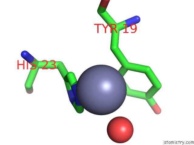
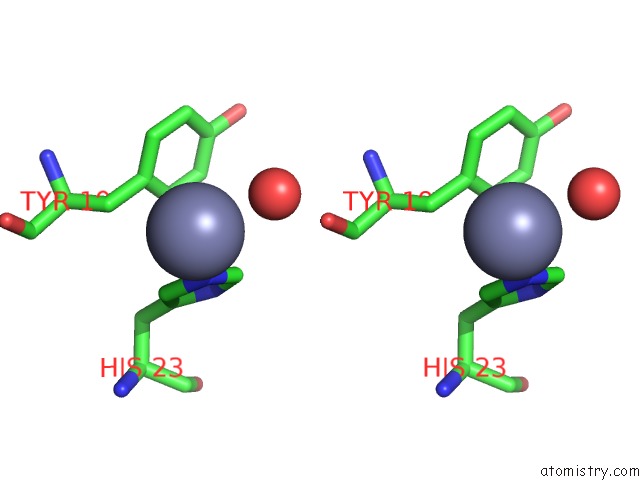
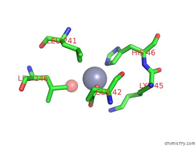
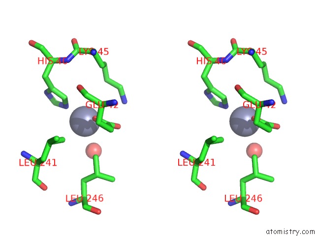
Zinc binding site 3 out of 4 in 3m3k
Go back to
Zinc binding site 3 out
of 4 in the Ligand Binding Domain (S1S2) of GLUA3 (Flop)
Mono view
Stereo pair view
Mono view
Stereo pair view
A full contact list of Zinc with other atoms in the Zn binding
site number 3 of Ligand Binding Domain (S1S2) of GLUA3 (Flop) within 5.0Å range:
|
Zinc binding site 4 out of 4 in 3m3k
Go back to
Zinc binding site 4 out
of 4 in the Ligand Binding Domain (S1S2) of GLUA3 (Flop)
Mono view
Stereo pair view
Mono view
Stereo pair view
A full contact list of Zinc with other atoms in the Zn binding
site number 4 of Ligand Binding Domain (S1S2) of GLUA3 (Flop) within 5.0Å range:
|
Reference:
A.H.Ahmed,
C.P.Ptak,
R.E.Oswald.
Molecular Mechanism of Flop Selectivity and Subsite Recognition For An Ampa Receptor Allosteric Modulator: Structures of GLUA2 and GLUA3 in Complexes with Pepa. Biochemistry V. 49 2843 2010.
ISSN: ISSN 0006-2960
PubMed: 20199107
DOI: 10.1021/BI1000678
Page generated: Sat Oct 26 09:05:15 2024
ISSN: ISSN 0006-2960
PubMed: 20199107
DOI: 10.1021/BI1000678
Last articles
Zn in 9MJ5Zn in 9HNW
Zn in 9G0L
Zn in 9FNE
Zn in 9DZN
Zn in 9E0I
Zn in 9D32
Zn in 9DAK
Zn in 8ZXC
Zn in 8ZUF