Zinc »
PDB 3lta-3m1m »
3m1j »
Zinc in PDB 3m1j: The Crystal Structure of A Nami A-Carbonic Anhydrase II Adduct Discloses the Mode of Action of This Novel Anticancer Metallodrug
Enzymatic activity of The Crystal Structure of A Nami A-Carbonic Anhydrase II Adduct Discloses the Mode of Action of This Novel Anticancer Metallodrug
All present enzymatic activity of The Crystal Structure of A Nami A-Carbonic Anhydrase II Adduct Discloses the Mode of Action of This Novel Anticancer Metallodrug:
4.2.1.1;
4.2.1.1;
Protein crystallography data
The structure of The Crystal Structure of A Nami A-Carbonic Anhydrase II Adduct Discloses the Mode of Action of This Novel Anticancer Metallodrug, PDB code: 3m1j
was solved by
C.Temperini,
L.Messori,
with X-Ray Crystallography technique. A brief refinement statistics is given in the table below:
| Resolution Low / High (Å) | 10.87 / 1.80 |
| Space group | P 1 21 1 |
| Cell size a, b, c (Å), α, β, γ (°) | 42.100, 41.550, 72.290, 90.00, 104.51, 90.00 |
| R / Rfree (%) | 19.3 / 23.5 |
Other elements in 3m1j:
The structure of The Crystal Structure of A Nami A-Carbonic Anhydrase II Adduct Discloses the Mode of Action of This Novel Anticancer Metallodrug also contains other interesting chemical elements:
| Ruthenium | (Ru) | 1 atom |
| Mercury | (Hg) | 1 atom |
Zinc Binding Sites:
The binding sites of Zinc atom in the The Crystal Structure of A Nami A-Carbonic Anhydrase II Adduct Discloses the Mode of Action of This Novel Anticancer Metallodrug
(pdb code 3m1j). This binding sites where shown within
5.0 Angstroms radius around Zinc atom.
In total only one binding site of Zinc was determined in the The Crystal Structure of A Nami A-Carbonic Anhydrase II Adduct Discloses the Mode of Action of This Novel Anticancer Metallodrug, PDB code: 3m1j:
In total only one binding site of Zinc was determined in the The Crystal Structure of A Nami A-Carbonic Anhydrase II Adduct Discloses the Mode of Action of This Novel Anticancer Metallodrug, PDB code: 3m1j:
Zinc binding site 1 out of 1 in 3m1j
Go back to
Zinc binding site 1 out
of 1 in the The Crystal Structure of A Nami A-Carbonic Anhydrase II Adduct Discloses the Mode of Action of This Novel Anticancer Metallodrug

Mono view
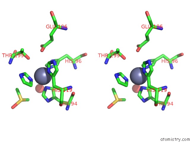
Stereo pair view
Mono view
Stereo pair view
A full contact list of Zinc with other atoms in the Zn binding
site number 1 of The Crystal Structure of A Nami A-Carbonic Anhydrase II Adduct Discloses the Mode of Action of This Novel Anticancer Metallodrug within 5.0Å range:
|
Reference:
A.Casini,
C.Temperini,
C.Gabbiani,
C.T.Supuran,
L.Messori.
The X-Ray Structure of the Adduct Between Nami-A and Carbonic Anhydrase Provides Insights Into the Reactivity of This Metallodrug with Proteins Chemmedchem V. 5 1989 2010.
ISSN: ISSN 1860-7179
PubMed: 20931644
DOI: 10.1002/CMDC.201000331
Page generated: Sat Oct 26 09:00:45 2024
ISSN: ISSN 1860-7179
PubMed: 20931644
DOI: 10.1002/CMDC.201000331
Last articles
Zn in 9MJ5Zn in 9HNW
Zn in 9G0L
Zn in 9FNE
Zn in 9DZN
Zn in 9E0I
Zn in 9D32
Zn in 9DAK
Zn in 8ZXC
Zn in 8ZUF