Zinc »
PDB 3lju-3lt9 »
3ls9 »
Zinc in PDB 3ls9: Crystal Structure of Atrazine Chlorohydrolase Trzn From Arthrobacter Aurescens TC1 Complexed with Zinc
Protein crystallography data
The structure of Crystal Structure of Atrazine Chlorohydrolase Trzn From Arthrobacter Aurescens TC1 Complexed with Zinc, PDB code: 3ls9
was solved by
A.A.Fedorov,
E.V.Fedorov,
J.Seffernick,
L.P.Wackett,
S.C.Almo,
with X-Ray Crystallography technique. A brief refinement statistics is given in the table below:
| Resolution Low / High (Å) | 31.86 / 1.40 |
| Space group | P 1 21 1 |
| Cell size a, b, c (Å), α, β, γ (°) | 57.552, 102.616, 80.553, 90.00, 104.75, 90.00 |
| R / Rfree (%) | 16.9 / 18.9 |
Zinc Binding Sites:
The binding sites of Zinc atom in the Crystal Structure of Atrazine Chlorohydrolase Trzn From Arthrobacter Aurescens TC1 Complexed with Zinc
(pdb code 3ls9). This binding sites where shown within
5.0 Angstroms radius around Zinc atom.
In total 2 binding sites of Zinc where determined in the Crystal Structure of Atrazine Chlorohydrolase Trzn From Arthrobacter Aurescens TC1 Complexed with Zinc, PDB code: 3ls9:
Jump to Zinc binding site number: 1; 2;
In total 2 binding sites of Zinc where determined in the Crystal Structure of Atrazine Chlorohydrolase Trzn From Arthrobacter Aurescens TC1 Complexed with Zinc, PDB code: 3ls9:
Jump to Zinc binding site number: 1; 2;
Zinc binding site 1 out of 2 in 3ls9
Go back to
Zinc binding site 1 out
of 2 in the Crystal Structure of Atrazine Chlorohydrolase Trzn From Arthrobacter Aurescens TC1 Complexed with Zinc
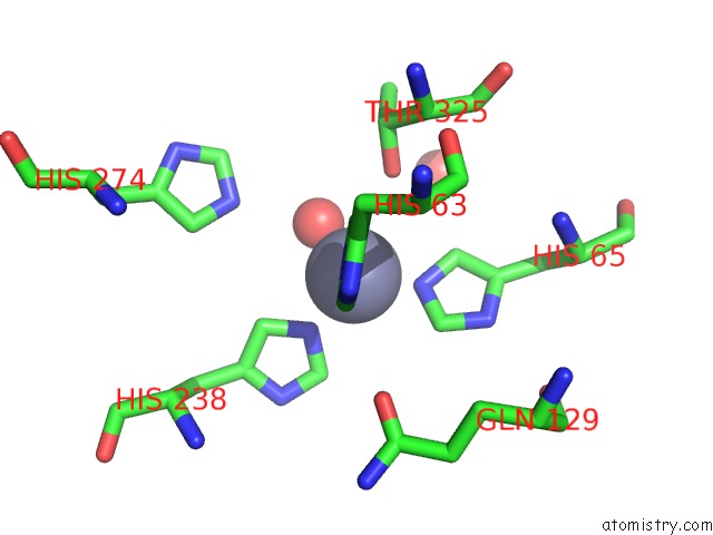
Mono view
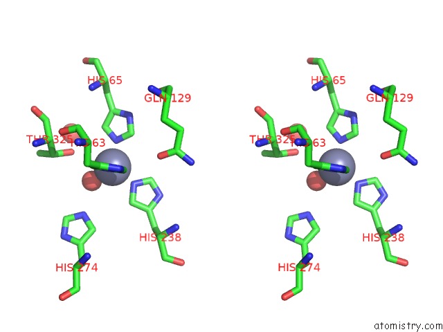
Stereo pair view
Mono view
Stereo pair view
A full contact list of Zinc with other atoms in the Zn binding
site number 1 of Crystal Structure of Atrazine Chlorohydrolase Trzn From Arthrobacter Aurescens TC1 Complexed with Zinc within 5.0Å range:
|
Zinc binding site 2 out of 2 in 3ls9
Go back to
Zinc binding site 2 out
of 2 in the Crystal Structure of Atrazine Chlorohydrolase Trzn From Arthrobacter Aurescens TC1 Complexed with Zinc
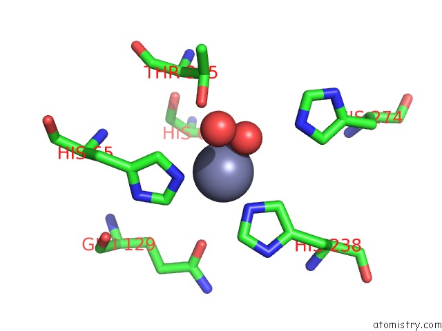
Mono view
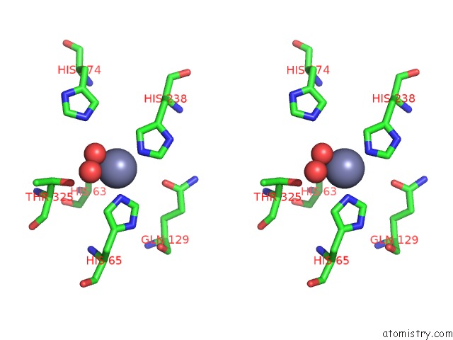
Stereo pair view
Mono view
Stereo pair view
A full contact list of Zinc with other atoms in the Zn binding
site number 2 of Crystal Structure of Atrazine Chlorohydrolase Trzn From Arthrobacter Aurescens TC1 Complexed with Zinc within 5.0Å range:
|
Reference:
A.A.Fedorov,
E.V.Fedorov,
J.Seffernick,
L.P.Wackett,
S.C.Almo.
Crystal Structure of Atrazine Chlorohydrolase Trzn From Arthrobacter Aurescens TC1 Complexed with Zinc To Be Published.
Page generated: Sat Oct 26 08:49:14 2024
Last articles
K in 1KEEK in 1M1K
K in 1LRT
K in 1M0Q
K in 1M0P
K in 1M0O
K in 1M0N
K in 1LWG
K in 1LW9
K in 1LVG