Zinc »
PDB 3lju-3lt9 »
3ls1 »
Zinc in PDB 3ls1: Crystal Structure of Cyanobacterial Psbq From Synechocystis Sp. Pcc 6803 Complexed with ZN2+
Protein crystallography data
The structure of Crystal Structure of Cyanobacterial Psbq From Synechocystis Sp. Pcc 6803 Complexed with ZN2+, PDB code: 3ls1
was solved by
S.A.Jackson,
R.D.Fagerlund,
S.M.Wilbanks,
J.J.Eaton-Rye,
with X-Ray Crystallography technique. A brief refinement statistics is given in the table below:
| Resolution Low / High (Å) | 21.76 / 1.85 |
| Space group | I 1 2 1 |
| Cell size a, b, c (Å), α, β, γ (°) | 92.771, 29.030, 99.503, 90.00, 97.77, 90.00 |
| R / Rfree (%) | 17.9 / 21.8 |
Zinc Binding Sites:
The binding sites of Zinc atom in the Crystal Structure of Cyanobacterial Psbq From Synechocystis Sp. Pcc 6803 Complexed with ZN2+
(pdb code 3ls1). This binding sites where shown within
5.0 Angstroms radius around Zinc atom.
In total 4 binding sites of Zinc where determined in the Crystal Structure of Cyanobacterial Psbq From Synechocystis Sp. Pcc 6803 Complexed with ZN2+, PDB code: 3ls1:
Jump to Zinc binding site number: 1; 2; 3; 4;
In total 4 binding sites of Zinc where determined in the Crystal Structure of Cyanobacterial Psbq From Synechocystis Sp. Pcc 6803 Complexed with ZN2+, PDB code: 3ls1:
Jump to Zinc binding site number: 1; 2; 3; 4;
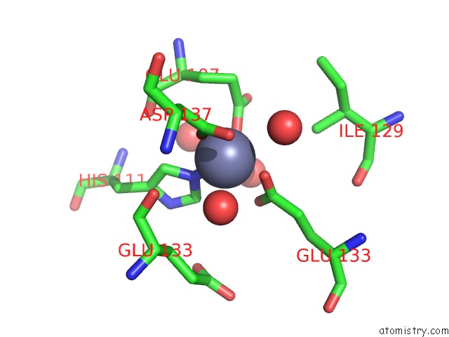
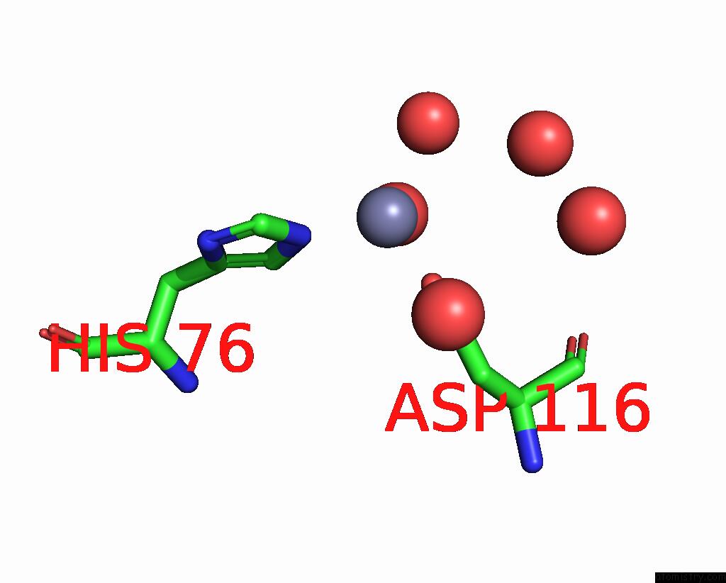
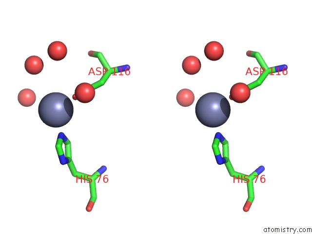
Zinc binding site 1 out of 4 in 3ls1
Go back to
Zinc binding site 1 out
of 4 in the Crystal Structure of Cyanobacterial Psbq From Synechocystis Sp. Pcc 6803 Complexed with ZN2+
Mono view
Stereo pair view
Mono view
Stereo pair view
A full contact list of Zinc with other atoms in the Zn binding
site number 1 of Crystal Structure of Cyanobacterial Psbq From Synechocystis Sp. Pcc 6803 Complexed with ZN2+ within 5.0Å range:
|
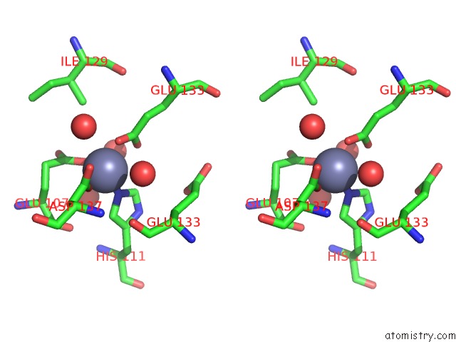
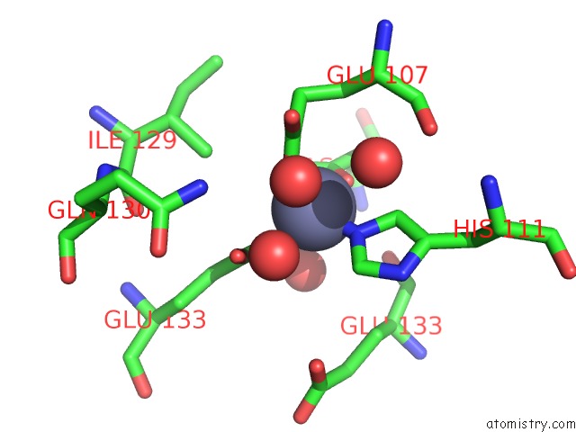
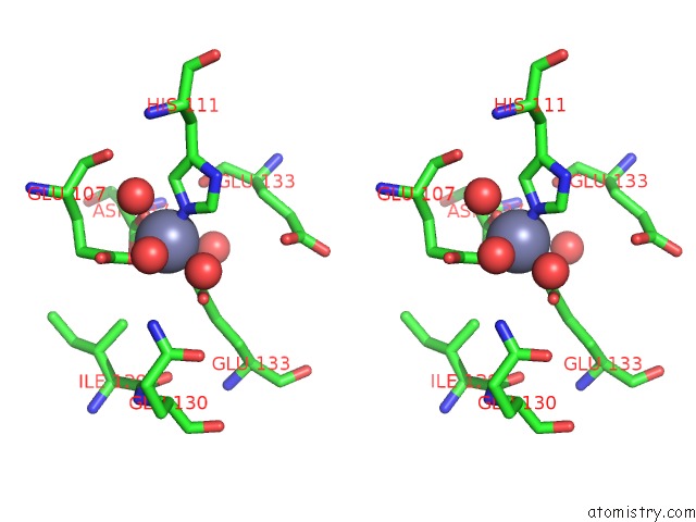
Zinc binding site 2 out of 4 in 3ls1
Go back to
Zinc binding site 2 out
of 4 in the Crystal Structure of Cyanobacterial Psbq From Synechocystis Sp. Pcc 6803 Complexed with ZN2+
Mono view
Stereo pair view
Mono view
Stereo pair view
A full contact list of Zinc with other atoms in the Zn binding
site number 2 of Crystal Structure of Cyanobacterial Psbq From Synechocystis Sp. Pcc 6803 Complexed with ZN2+ within 5.0Å range:
|
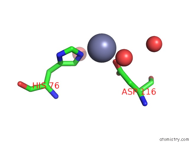
Zinc binding site 3 out of 4 in 3ls1
Go back to
Zinc binding site 3 out
of 4 in the Crystal Structure of Cyanobacterial Psbq From Synechocystis Sp. Pcc 6803 Complexed with ZN2+
Mono view
Stereo pair view
Mono view
Stereo pair view
A full contact list of Zinc with other atoms in the Zn binding
site number 3 of Crystal Structure of Cyanobacterial Psbq From Synechocystis Sp. Pcc 6803 Complexed with ZN2+ within 5.0Å range:
|
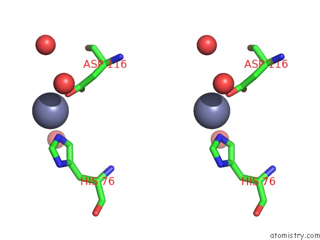
Zinc binding site 4 out of 4 in 3ls1
Go back to
Zinc binding site 4 out
of 4 in the Crystal Structure of Cyanobacterial Psbq From Synechocystis Sp. Pcc 6803 Complexed with ZN2+
Mono view
Stereo pair view
Mono view
Stereo pair view
A full contact list of Zinc with other atoms in the Zn binding
site number 4 of Crystal Structure of Cyanobacterial Psbq From Synechocystis Sp. Pcc 6803 Complexed with ZN2+ within 5.0Å range:
|
Reference:
S.A.Jackson,
R.D.Fagerlund,
S.M.Wilbanks,
J.J.Eaton-Rye.
Crystal Structure of Psbq From Synechocystis Sp. Pcc 6803 at 1.8 A: Implications For Binding and Function in Cyanobacterial Photosystem II Biochemistry V. 49 2765 2010.
ISSN: ISSN 0006-2960
PubMed: 20210304
DOI: 10.1021/BI100217H
Page generated: Sat Oct 26 08:48:13 2024
ISSN: ISSN 0006-2960
PubMed: 20210304
DOI: 10.1021/BI100217H
Last articles
Zn in 9MJ5Zn in 9HNW
Zn in 9G0L
Zn in 9FNE
Zn in 9DZN
Zn in 9E0I
Zn in 9D32
Zn in 9DAK
Zn in 8ZXC
Zn in 8ZUF