Zinc »
PDB 3ljz-3lta »
3lqb »
Zinc in PDB 3lqb: Crystal Structure of the Hatching Enzyme ZHE1 From the Zebrafish Danio Rerio
Protein crystallography data
The structure of Crystal Structure of the Hatching Enzyme ZHE1 From the Zebrafish Danio Rerio, PDB code: 3lqb
was solved by
M.Tanokura,
A.Okada,
K.Nagata,
S.Yasumasu,
J.Ohtsuka,
I.Iuchi,
with X-Ray Crystallography technique. A brief refinement statistics is given in the table below:
| Resolution Low / High (Å) | 14.98 / 1.10 |
| Space group | P 21 21 21 |
| Cell size a, b, c (Å), α, β, γ (°) | 32.970, 62.680, 87.340, 90.00, 90.00, 90.00 |
| R / Rfree (%) | 16.9 / 18.7 |
Zinc Binding Sites:
The binding sites of Zinc atom in the Crystal Structure of the Hatching Enzyme ZHE1 From the Zebrafish Danio Rerio
(pdb code 3lqb). This binding sites where shown within
5.0 Angstroms radius around Zinc atom.
In total only one binding site of Zinc was determined in the Crystal Structure of the Hatching Enzyme ZHE1 From the Zebrafish Danio Rerio, PDB code: 3lqb:
In total only one binding site of Zinc was determined in the Crystal Structure of the Hatching Enzyme ZHE1 From the Zebrafish Danio Rerio, PDB code: 3lqb:
Zinc binding site 1 out of 1 in 3lqb
Go back to
Zinc binding site 1 out
of 1 in the Crystal Structure of the Hatching Enzyme ZHE1 From the Zebrafish Danio Rerio

Mono view
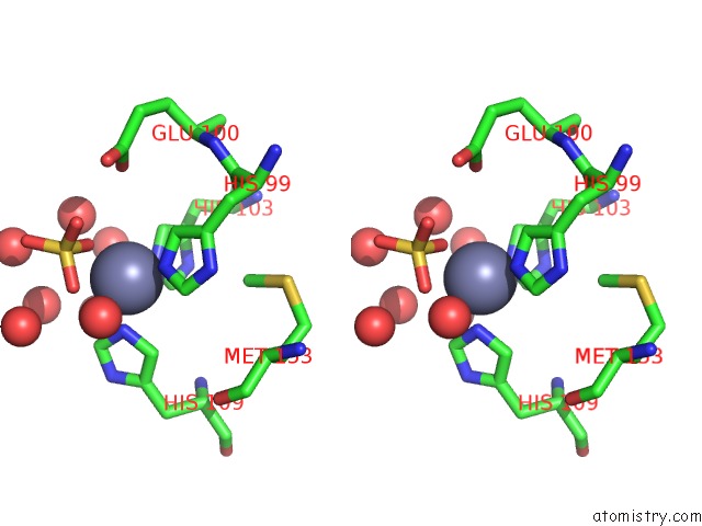
Stereo pair view
Mono view
Stereo pair view
A full contact list of Zinc with other atoms in the Zn binding
site number 1 of Crystal Structure of the Hatching Enzyme ZHE1 From the Zebrafish Danio Rerio within 5.0Å range:
|
Reference:
A.Okada,
K.Sano,
K.Nagata,
S.Yasumasu,
J.Ohtsuka,
A.Yamamura,
K.Kubota,
I.Iuchi,
M.Tanokura.
Crystal Structure of Zebrafish Hatching Enzyme 1 From the Zebrafish Danio Rerio J.Mol.Biol. V. 402 865 2010.
ISSN: ISSN 0022-2836
PubMed: 20727360
DOI: 10.1016/J.JMB.2010.08.023
Page generated: Wed Aug 20 11:25:39 2025
ISSN: ISSN 0022-2836
PubMed: 20727360
DOI: 10.1016/J.JMB.2010.08.023
Last articles
Zn in 3S77Zn in 3S45
Zn in 3S76
Zn in 3S75
Zn in 3S74
Zn in 3S73
Zn in 3S72
Zn in 3S71
Zn in 3S5M
Zn in 3S5K