Zinc »
PDB 3l2v-3ljt »
3l7t »
Zinc in PDB 3l7t: Crystal Structure of Smu.1112C
Protein crystallography data
The structure of Crystal Structure of Smu.1112C, PDB code: 3l7t
was solved by
X.-X.Fan,
K.-T.Wang,
X.-D.Su,
with X-Ray Crystallography technique. A brief refinement statistics is given in the table below:
| Resolution Low / High (Å) | 28.83 / 1.80 |
| Space group | P 1 21 1 |
| Cell size a, b, c (Å), α, β, γ (°) | 47.603, 72.577, 86.851, 90.00, 94.45, 90.00 |
| R / Rfree (%) | 22.2 / 26.2 |
Zinc Binding Sites:
The binding sites of Zinc atom in the Crystal Structure of Smu.1112C
(pdb code 3l7t). This binding sites where shown within
5.0 Angstroms radius around Zinc atom.
In total 2 binding sites of Zinc where determined in the Crystal Structure of Smu.1112C, PDB code: 3l7t:
Jump to Zinc binding site number: 1; 2;
In total 2 binding sites of Zinc where determined in the Crystal Structure of Smu.1112C, PDB code: 3l7t:
Jump to Zinc binding site number: 1; 2;
Zinc binding site 1 out of 2 in 3l7t
Go back to
Zinc binding site 1 out
of 2 in the Crystal Structure of Smu.1112C
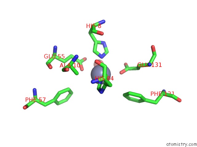
Mono view
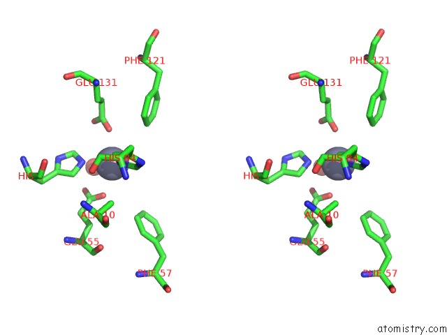
Stereo pair view
Mono view
Stereo pair view
A full contact list of Zinc with other atoms in the Zn binding
site number 1 of Crystal Structure of Smu.1112C within 5.0Å range:
|
Zinc binding site 2 out of 2 in 3l7t
Go back to
Zinc binding site 2 out
of 2 in the Crystal Structure of Smu.1112C
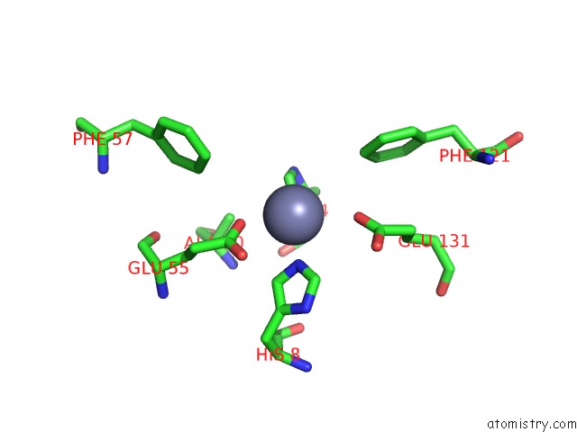
Mono view
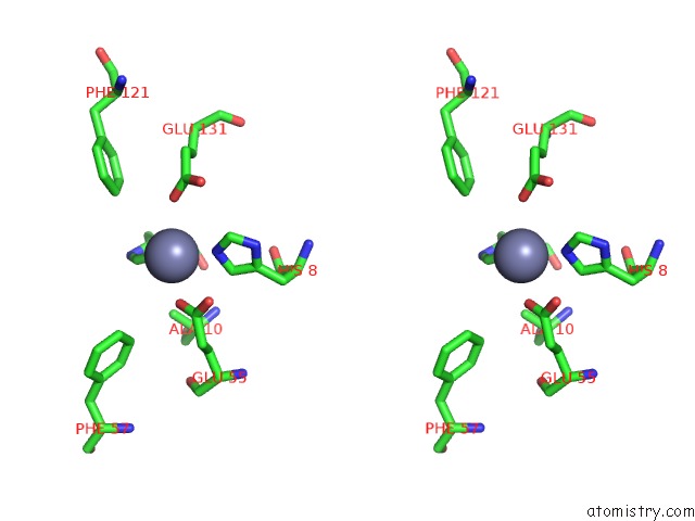
Stereo pair view
Mono view
Stereo pair view
A full contact list of Zinc with other atoms in the Zn binding
site number 2 of Crystal Structure of Smu.1112C within 5.0Å range:
|
Reference:
X.-X.Fan,
K.-T.Wang,
X.-D.Su.
Crystal Structure of Smu.1112C To Be Published.
Page generated: Sat Oct 26 08:25:12 2024
Last articles
As in 3FPCAs in 3FRG
As in 3FPL
As in 3FKG
As in 3FM4
As in 3FMU
As in 3ET6
As in 3ENZ
As in 3FJU
As in 3FCU