Zinc »
PDB 3kry-3l2v »
3l11 »
Zinc in PDB 3l11: Crystal Structure of the Ring Domain of RNF168
Protein crystallography data
The structure of Crystal Structure of the Ring Domain of RNF168, PDB code: 3l11
was solved by
D.Neculai,
L.Yermekbayeva,
L.Crombet,
J.Weigelt,
C.Bountra,
A.M.Edwards,
C.H.Arrowsmith,
A.Bochkarev,
S.Dhe-Paganon,
with X-Ray Crystallography technique. A brief refinement statistics is given in the table below:
| Resolution Low / High (Å) | 45.49 / 2.12 |
| Space group | P 43 21 2 |
| Cell size a, b, c (Å), α, β, γ (°) | 49.706, 49.706, 110.149, 90.00, 90.00, 90.00 |
| R / Rfree (%) | 18.7 / 21.5 |
Zinc Binding Sites:
The binding sites of Zinc atom in the Crystal Structure of the Ring Domain of RNF168
(pdb code 3l11). This binding sites where shown within
5.0 Angstroms radius around Zinc atom.
In total 2 binding sites of Zinc where determined in the Crystal Structure of the Ring Domain of RNF168, PDB code: 3l11:
Jump to Zinc binding site number: 1; 2;
In total 2 binding sites of Zinc where determined in the Crystal Structure of the Ring Domain of RNF168, PDB code: 3l11:
Jump to Zinc binding site number: 1; 2;
Zinc binding site 1 out of 2 in 3l11
Go back to
Zinc binding site 1 out
of 2 in the Crystal Structure of the Ring Domain of RNF168
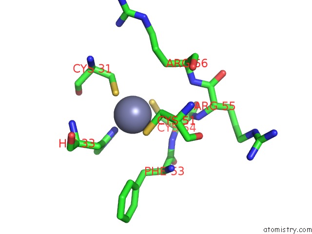
Mono view
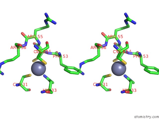
Stereo pair view
Mono view
Stereo pair view
A full contact list of Zinc with other atoms in the Zn binding
site number 1 of Crystal Structure of the Ring Domain of RNF168 within 5.0Å range:
|
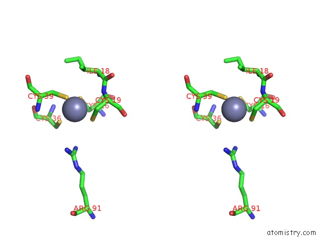
Zinc binding site 2 out of 2 in 3l11
Go back to
Zinc binding site 2 out
of 2 in the Crystal Structure of the Ring Domain of RNF168
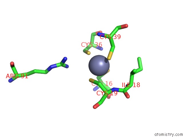
Mono view
Stereo pair view
Mono view
Stereo pair view
A full contact list of Zinc with other atoms in the Zn binding
site number 2 of Crystal Structure of the Ring Domain of RNF168 within 5.0Å range:
|
Reference:
S.J.Campbell,
R.A.Edwards,
C.C.Leung,
D.Neculai,
C.D.Hodge,
S.Dhe-Paganon,
J.N.Glover.
Molecular Insights Into the Function of Ring Finger (Rnf)-Containing Proteins HRNF8 and HRNF168 in UBC13/MMS2-Dependent Ubiquitylation. J.Biol.Chem. V. 287 23900 2012.
ISSN: ISSN 0021-9258
PubMed: 22589545
DOI: 10.1074/JBC.M112.359653
Page generated: Wed Aug 20 11:11:44 2025
ISSN: ISSN 0021-9258
PubMed: 22589545
DOI: 10.1074/JBC.M112.359653
Last articles
Zn in 3SASZn in 3S6L
Zn in 3S9C
Zn in 3SAR
Zn in 3SAP
Zn in 3S9T
Zn in 3S7J
Zn in 3S8P
Zn in 3S8X
Zn in 3S7F