Zinc »
PDB 3kry-3l2v »
3l0a »
Zinc in PDB 3l0a: Crystal Structure of Putative Exonuclease (RER070207002219) From Eubacterium Rectale at 2.19 A Resolution
Protein crystallography data
The structure of Crystal Structure of Putative Exonuclease (RER070207002219) From Eubacterium Rectale at 2.19 A Resolution, PDB code: 3l0a
was solved by
Joint Center For Structural Genomics (Jcsg),
with X-Ray Crystallography technique. A brief refinement statistics is given in the table below:
| Resolution Low / High (Å) | 44.11 / 2.19 |
| Space group | P 63 |
| Cell size a, b, c (Å), α, β, γ (°) | 88.248, 88.248, 69.350, 90.00, 90.00, 120.00 |
| R / Rfree (%) | 20.8 / 25 |
Zinc Binding Sites:
The binding sites of Zinc atom in the Crystal Structure of Putative Exonuclease (RER070207002219) From Eubacterium Rectale at 2.19 A Resolution
(pdb code 3l0a). This binding sites where shown within
5.0 Angstroms radius around Zinc atom.
In total only one binding site of Zinc was determined in the Crystal Structure of Putative Exonuclease (RER070207002219) From Eubacterium Rectale at 2.19 A Resolution, PDB code: 3l0a:
In total only one binding site of Zinc was determined in the Crystal Structure of Putative Exonuclease (RER070207002219) From Eubacterium Rectale at 2.19 A Resolution, PDB code: 3l0a:
Zinc binding site 1 out of 1 in 3l0a
Go back to
Zinc binding site 1 out
of 1 in the Crystal Structure of Putative Exonuclease (RER070207002219) From Eubacterium Rectale at 2.19 A Resolution
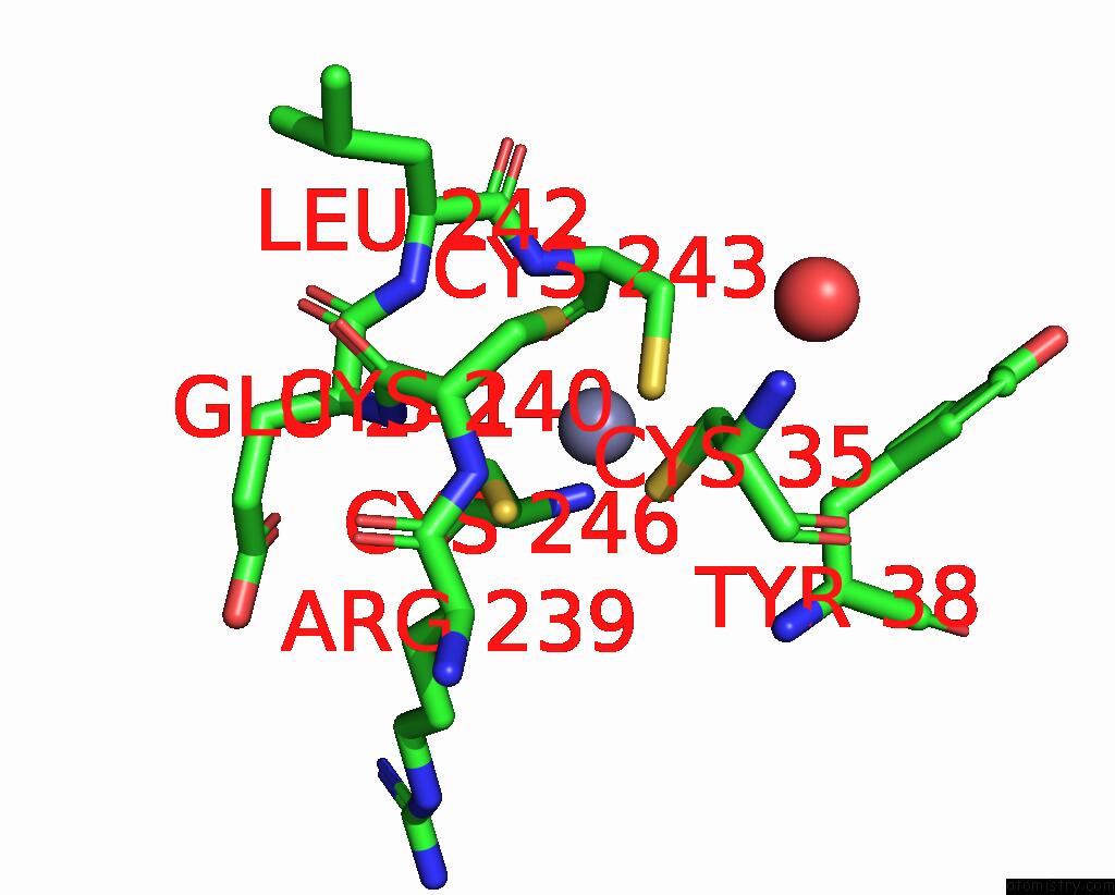
Mono view
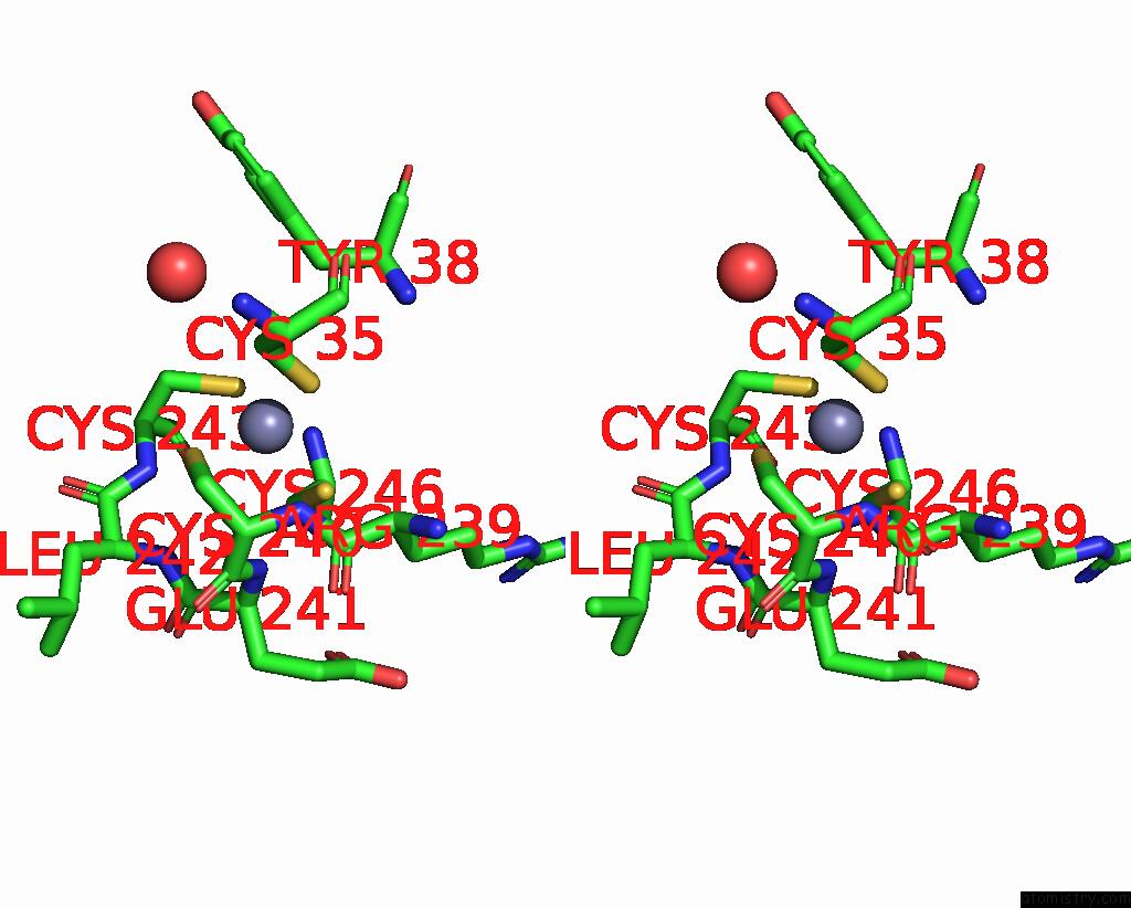
Stereo pair view
Mono view
Stereo pair view
A full contact list of Zinc with other atoms in the Zn binding
site number 1 of Crystal Structure of Putative Exonuclease (RER070207002219) From Eubacterium Rectale at 2.19 A Resolution within 5.0Å range:
|
Reference:
Joint Center For Structural Genomics (Jcsg),
Joint Center For Structural Genomics (Jcsg).
N/A N/A.
Page generated: Wed Aug 20 11:11:29 2025
Last articles
Zn in 4GD4Zn in 4GEL
Zn in 4GD0
Zn in 4GCX
Zn in 4GC3
Zn in 4GBM
Zn in 4GCW
Zn in 4GBN
Zn in 4GBD
Zn in 4GBL