Zinc »
PDB 3iqx-3jr3 »
3ivt »
Zinc in PDB 3ivt: Homocitrate Synthase LYS4 Bound to 2-Og
Enzymatic activity of Homocitrate Synthase LYS4 Bound to 2-Og
All present enzymatic activity of Homocitrate Synthase LYS4 Bound to 2-Og:
2.3.3.14;
2.3.3.14;
Protein crystallography data
The structure of Homocitrate Synthase LYS4 Bound to 2-Og, PDB code: 3ivt
was solved by
S.L.Bulfer,
E.M.Scott,
J.-F.Couture,
L.Pillus,
R.C.Trievel,
with X-Ray Crystallography technique. A brief refinement statistics is given in the table below:
| Resolution Low / High (Å) | 39.47 / 2.67 |
| Space group | P 62 |
| Cell size a, b, c (Å), α, β, γ (°) | 133.731, 133.731, 125.904, 90.00, 90.00, 120.00 |
| R / Rfree (%) | 16.5 / 20.6 |
Other elements in 3ivt:
The structure of Homocitrate Synthase LYS4 Bound to 2-Og also contains other interesting chemical elements:
| Sodium | (Na) | 2 atoms |
Zinc Binding Sites:
The binding sites of Zinc atom in the Homocitrate Synthase LYS4 Bound to 2-Og
(pdb code 3ivt). This binding sites where shown within
5.0 Angstroms radius around Zinc atom.
In total 2 binding sites of Zinc where determined in the Homocitrate Synthase LYS4 Bound to 2-Og, PDB code: 3ivt:
Jump to Zinc binding site number: 1; 2;
In total 2 binding sites of Zinc where determined in the Homocitrate Synthase LYS4 Bound to 2-Og, PDB code: 3ivt:
Jump to Zinc binding site number: 1; 2;
Zinc binding site 1 out of 2 in 3ivt
Go back to
Zinc binding site 1 out
of 2 in the Homocitrate Synthase LYS4 Bound to 2-Og
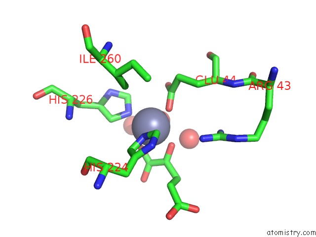
Mono view
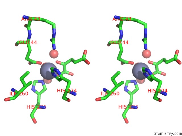
Stereo pair view
Mono view
Stereo pair view
A full contact list of Zinc with other atoms in the Zn binding
site number 1 of Homocitrate Synthase LYS4 Bound to 2-Og within 5.0Å range:
|
Zinc binding site 2 out of 2 in 3ivt
Go back to
Zinc binding site 2 out
of 2 in the Homocitrate Synthase LYS4 Bound to 2-Og
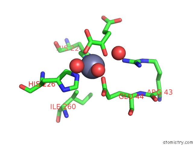
Mono view
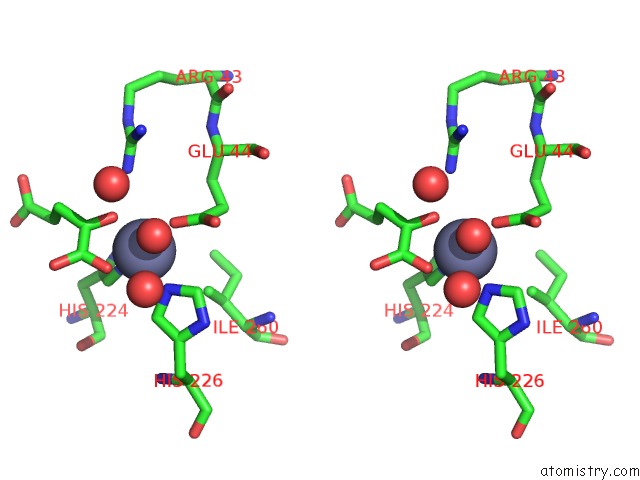
Stereo pair view
Mono view
Stereo pair view
A full contact list of Zinc with other atoms in the Zn binding
site number 2 of Homocitrate Synthase LYS4 Bound to 2-Og within 5.0Å range:
|
Reference:
S.L.Bulfer,
E.M.Scott,
J.F.Couture,
L.Pillus,
R.C.Trievel.
Crystal Structure and Functional Analysis of Homocitrate Synthase, An Essential Enzyme in Lysine Biosynthesis. J.Biol.Chem. V. 284 35769 2009.
ISSN: ISSN 0021-9258
PubMed: 19776021
DOI: 10.1074/JBC.M109.046821
Page generated: Sat Oct 26 07:21:55 2024
ISSN: ISSN 0021-9258
PubMed: 19776021
DOI: 10.1074/JBC.M109.046821
Last articles
Zn in 9MJ5Zn in 9HNW
Zn in 9G0L
Zn in 9FNE
Zn in 9DZN
Zn in 9E0I
Zn in 9D32
Zn in 9DAK
Zn in 8ZXC
Zn in 8ZUF