Zinc »
PDB 3ifj-3iqx »
3ins »
Zinc in PDB 3ins: Structure of Insulin. Results of Joint Neutron and X-Ray Refinement
Protein crystallography data
The structure of Structure of Insulin. Results of Joint Neutron and X-Ray Refinement, PDB code: 3ins
was solved by
A.Wlodawer,
H.Savage,
with X-Ray Crystallography technique. A brief refinement statistics is given in the table below:
| Resolution Low / High (Å) | N/A / 1.50 |
| Space group | H 3 |
| Cell size a, b, c (Å), α, β, γ (°) | 82.500, 82.500, 34.000, 90.00, 90.00, 120.00 |
| R / Rfree (%) | n/a / n/a |
Zinc Binding Sites:
The binding sites of Zinc atom in the Structure of Insulin. Results of Joint Neutron and X-Ray Refinement
(pdb code 3ins). This binding sites where shown within
5.0 Angstroms radius around Zinc atom.
In total 2 binding sites of Zinc where determined in the Structure of Insulin. Results of Joint Neutron and X-Ray Refinement, PDB code: 3ins:
Jump to Zinc binding site number: 1; 2;
In total 2 binding sites of Zinc where determined in the Structure of Insulin. Results of Joint Neutron and X-Ray Refinement, PDB code: 3ins:
Jump to Zinc binding site number: 1; 2;
Zinc binding site 1 out of 2 in 3ins
Go back to
Zinc binding site 1 out
of 2 in the Structure of Insulin. Results of Joint Neutron and X-Ray Refinement
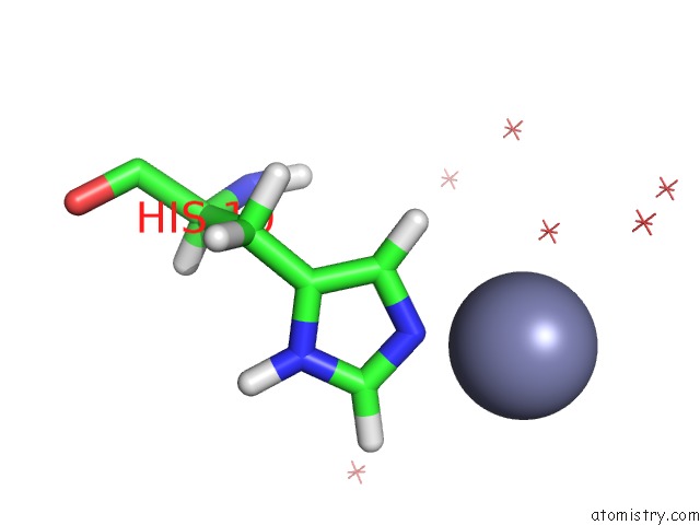
Mono view
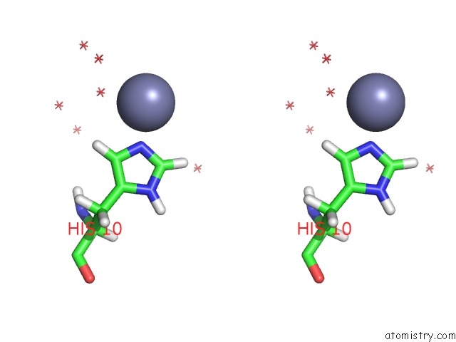
Stereo pair view
Mono view
Stereo pair view
A full contact list of Zinc with other atoms in the Zn binding
site number 1 of Structure of Insulin. Results of Joint Neutron and X-Ray Refinement within 5.0Å range:
|
Zinc binding site 2 out of 2 in 3ins
Go back to
Zinc binding site 2 out
of 2 in the Structure of Insulin. Results of Joint Neutron and X-Ray Refinement
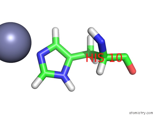
Mono view
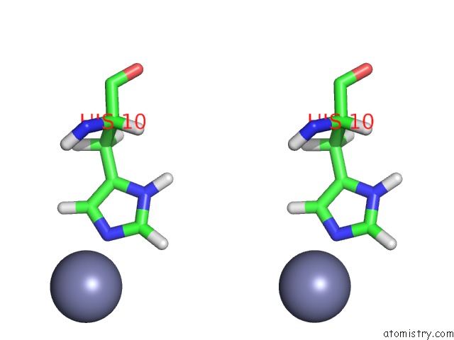
Stereo pair view
Mono view
Stereo pair view
A full contact list of Zinc with other atoms in the Zn binding
site number 2 of Structure of Insulin. Results of Joint Neutron and X-Ray Refinement within 5.0Å range:
|
Reference:
A.Wlodawer,
H.Savage,
G.Dodson.
Structure of Insulin: Results of Joint Neutron and X-Ray Refinement. Acta Crystallogr.,Sect.B V. 45 99 1989.
ISSN: ISSN 0108-7681
PubMed: 2695122
DOI: 10.1107/S0108768188011012
Page generated: Wed Aug 20 10:32:58 2025
ISSN: ISSN 0108-7681
PubMed: 2695122
DOI: 10.1107/S0108768188011012
Last articles
Zn in 4DXBZn in 4DWX
Zn in 4DWV
Zn in 4DWK
Zn in 4DVI
Zn in 4DWC
Zn in 4DV7
Zn in 4DV6
Zn in 4DV8
Zn in 4DV5