Zinc »
PDB 3ife-3iqw »
3ij6 »
Zinc in PDB 3ij6: Crystal Structure of An Uncharacterized Metal-Dependent Hydrolase From Lactobacillus Acidophilus
Protein crystallography data
The structure of Crystal Structure of An Uncharacterized Metal-Dependent Hydrolase From Lactobacillus Acidophilus, PDB code: 3ij6
was solved by
Y.Patskovsky,
R.Toro,
M.Dickey,
S.Chang,
J.M.Sauder,
F.M.Raushel,
S.K.Burley,
S.C.Almo,
New York Sgx Research Center For Structuralgenomics (Nysgxrc),
with X-Ray Crystallography technique. A brief refinement statistics is given in the table below:
| Resolution Low / High (Å) | 20.00 / 2.00 |
| Space group | C 1 2 1 |
| Cell size a, b, c (Å), α, β, γ (°) | 170.717, 96.433, 96.033, 90.00, 90.40, 90.00 |
| R / Rfree (%) | 22.1 / 26.8 |
Other elements in 3ij6:
The structure of Crystal Structure of An Uncharacterized Metal-Dependent Hydrolase From Lactobacillus Acidophilus also contains other interesting chemical elements:
| Sodium | (Na) | 4 atoms |
Zinc Binding Sites:
The binding sites of Zinc atom in the Crystal Structure of An Uncharacterized Metal-Dependent Hydrolase From Lactobacillus Acidophilus
(pdb code 3ij6). This binding sites where shown within
5.0 Angstroms radius around Zinc atom.
In total 4 binding sites of Zinc where determined in the Crystal Structure of An Uncharacterized Metal-Dependent Hydrolase From Lactobacillus Acidophilus, PDB code: 3ij6:
Jump to Zinc binding site number: 1; 2; 3; 4;
In total 4 binding sites of Zinc where determined in the Crystal Structure of An Uncharacterized Metal-Dependent Hydrolase From Lactobacillus Acidophilus, PDB code: 3ij6:
Jump to Zinc binding site number: 1; 2; 3; 4;
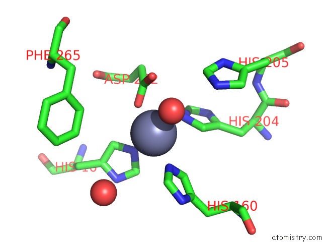
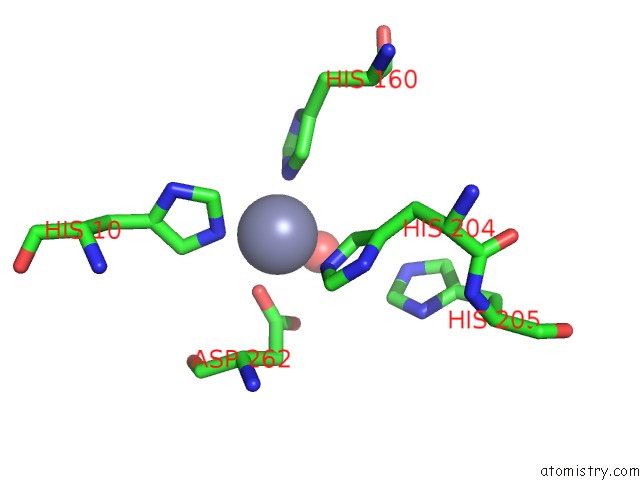
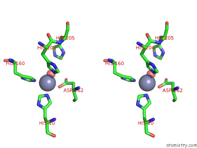
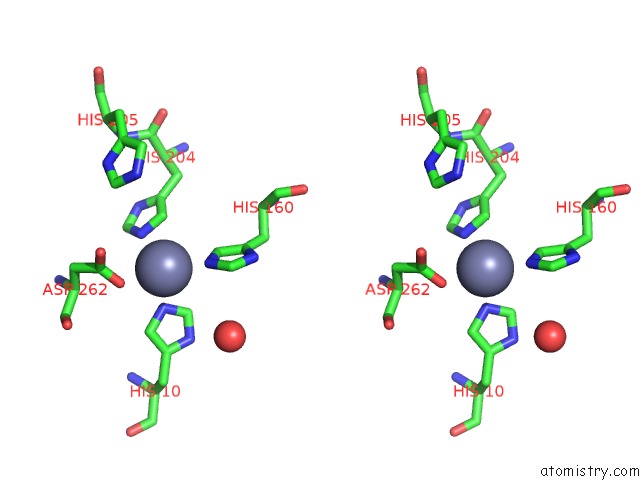
Zinc binding site 1 out of 4 in 3ij6
Go back to
Zinc binding site 1 out
of 4 in the Crystal Structure of An Uncharacterized Metal-Dependent Hydrolase From Lactobacillus Acidophilus

Mono view

Stereo pair view
Mono view
Stereo pair view
A full contact list of Zinc with other atoms in the Zn binding
site number 1 of Crystal Structure of An Uncharacterized Metal-Dependent Hydrolase From Lactobacillus Acidophilus within 5.0Å range:
|
Zinc binding site 2 out of 4 in 3ij6
Go back to
Zinc binding site 2 out
of 4 in the Crystal Structure of An Uncharacterized Metal-Dependent Hydrolase From Lactobacillus Acidophilus
Mono view

Stereo pair view
Mono view
Stereo pair view
A full contact list of Zinc with other atoms in the Zn binding
site number 2 of Crystal Structure of An Uncharacterized Metal-Dependent Hydrolase From Lactobacillus Acidophilus within 5.0Å range:
|
Zinc binding site 3 out of 4 in 3ij6
Go back to
Zinc binding site 3 out
of 4 in the Crystal Structure of An Uncharacterized Metal-Dependent Hydrolase From Lactobacillus Acidophilus
Mono view
Stereo pair view
Mono view
Stereo pair view
A full contact list of Zinc with other atoms in the Zn binding
site number 3 of Crystal Structure of An Uncharacterized Metal-Dependent Hydrolase From Lactobacillus Acidophilus within 5.0Å range:
|
Zinc binding site 4 out of 4 in 3ij6
Go back to
Zinc binding site 4 out
of 4 in the Crystal Structure of An Uncharacterized Metal-Dependent Hydrolase From Lactobacillus Acidophilus

Mono view
Stereo pair view
Mono view
Stereo pair view
A full contact list of Zinc with other atoms in the Zn binding
site number 4 of Crystal Structure of An Uncharacterized Metal-Dependent Hydrolase From Lactobacillus Acidophilus within 5.0Å range:
|
Reference:
Y.Patskovsky,
R.Toro,
M.Dickey,
S.Chang,
J.M.Sauder,
F.M.Raushel,
S.K.Burley,
S.C.Almo.
Crystal Structure of An Uncharacterized Metal-Dependent Hydrolase From Lactobacillus Acidopphilus To Be Published.
Page generated: Sat Oct 26 07:06:30 2024
Last articles
Zn in 9MJ5Zn in 9HNW
Zn in 9G0L
Zn in 9FNE
Zn in 9DZN
Zn in 9E0I
Zn in 9D32
Zn in 9DAK
Zn in 8ZXC
Zn in 8ZUF