Zinc »
PDB 3i7g-3if1 »
3ic1 »
Zinc in PDB 3ic1: Crystal Structure of Zinc-Bound Succinyl-Diaminopimelate Desuccinylase From Haemophilus Influenzae
Enzymatic activity of Crystal Structure of Zinc-Bound Succinyl-Diaminopimelate Desuccinylase From Haemophilus Influenzae
All present enzymatic activity of Crystal Structure of Zinc-Bound Succinyl-Diaminopimelate Desuccinylase From Haemophilus Influenzae:
3.5.1.18;
3.5.1.18;
Protein crystallography data
The structure of Crystal Structure of Zinc-Bound Succinyl-Diaminopimelate Desuccinylase From Haemophilus Influenzae, PDB code: 3ic1
was solved by
B.P.Nocek,
D.M.Gillner,
R.C.Holz,
A.Joachimiak,
Midwest Center Forstructural Genomics (Mcsg),
with X-Ray Crystallography technique. A brief refinement statistics is given in the table below:
| Resolution Low / High (Å) | 40.00 / 2.30 |
| Space group | P 21 21 21 |
| Cell size a, b, c (Å), α, β, γ (°) | 44.697, 95.751, 185.426, 90.00, 90.00, 90.00 |
| R / Rfree (%) | 19.4 / 25 |
Zinc Binding Sites:
The binding sites of Zinc atom in the Crystal Structure of Zinc-Bound Succinyl-Diaminopimelate Desuccinylase From Haemophilus Influenzae
(pdb code 3ic1). This binding sites where shown within
5.0 Angstroms radius around Zinc atom.
In total 4 binding sites of Zinc where determined in the Crystal Structure of Zinc-Bound Succinyl-Diaminopimelate Desuccinylase From Haemophilus Influenzae, PDB code: 3ic1:
Jump to Zinc binding site number: 1; 2; 3; 4;
In total 4 binding sites of Zinc where determined in the Crystal Structure of Zinc-Bound Succinyl-Diaminopimelate Desuccinylase From Haemophilus Influenzae, PDB code: 3ic1:
Jump to Zinc binding site number: 1; 2; 3; 4;
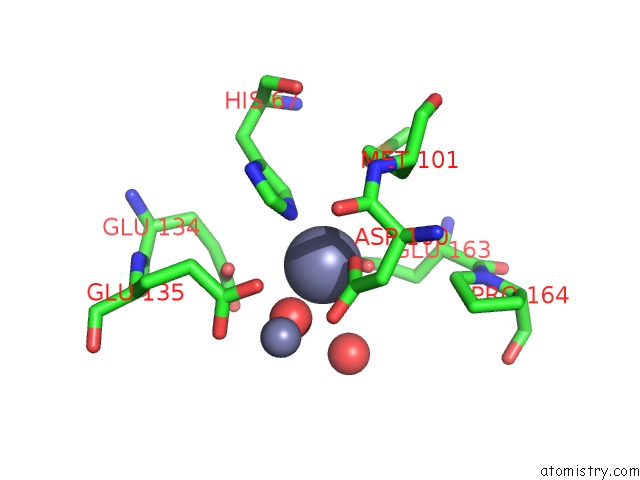
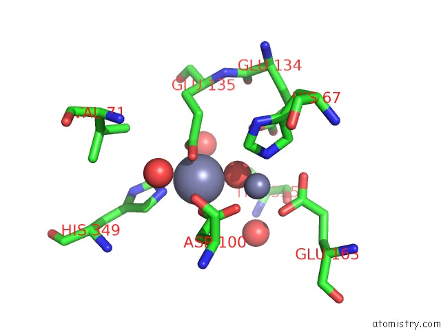
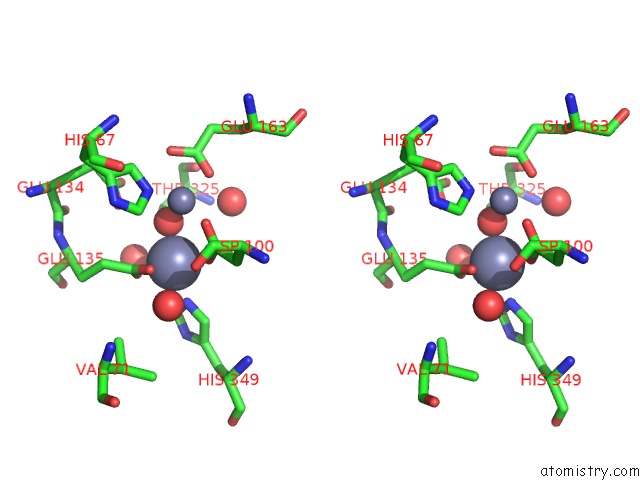
Zinc binding site 1 out of 4 in 3ic1
Go back to
Zinc binding site 1 out
of 4 in the Crystal Structure of Zinc-Bound Succinyl-Diaminopimelate Desuccinylase From Haemophilus Influenzae
Mono view
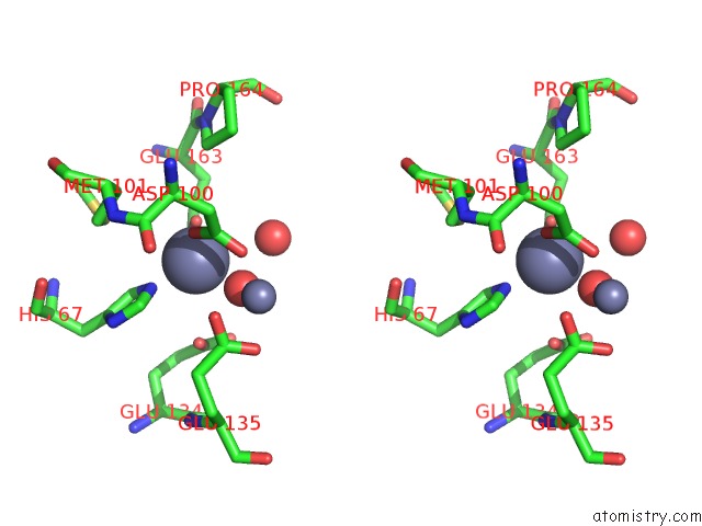
Stereo pair view
Mono view
Stereo pair view
A full contact list of Zinc with other atoms in the Zn binding
site number 1 of Crystal Structure of Zinc-Bound Succinyl-Diaminopimelate Desuccinylase From Haemophilus Influenzae within 5.0Å range:
|
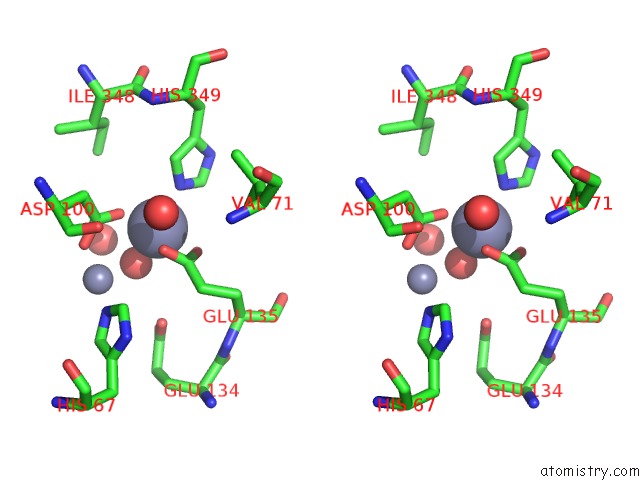
Zinc binding site 2 out of 4 in 3ic1
Go back to
Zinc binding site 2 out
of 4 in the Crystal Structure of Zinc-Bound Succinyl-Diaminopimelate Desuccinylase From Haemophilus Influenzae
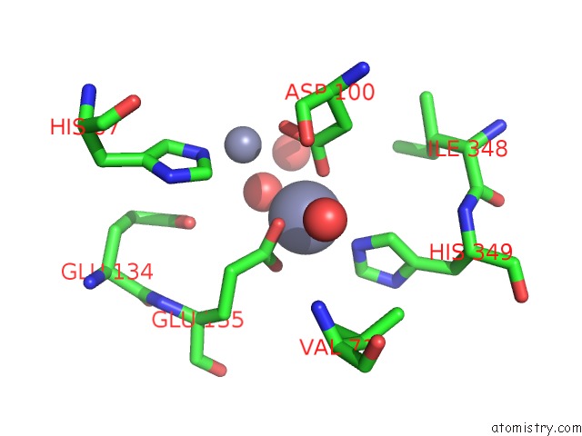
Mono view
Stereo pair view
Mono view
Stereo pair view
A full contact list of Zinc with other atoms in the Zn binding
site number 2 of Crystal Structure of Zinc-Bound Succinyl-Diaminopimelate Desuccinylase From Haemophilus Influenzae within 5.0Å range:
|
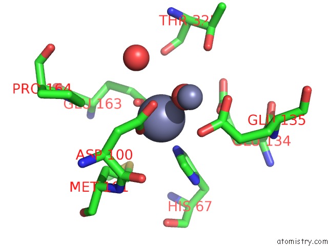
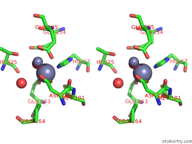
Zinc binding site 3 out of 4 in 3ic1
Go back to
Zinc binding site 3 out
of 4 in the Crystal Structure of Zinc-Bound Succinyl-Diaminopimelate Desuccinylase From Haemophilus Influenzae
Mono view
Stereo pair view
Mono view
Stereo pair view
A full contact list of Zinc with other atoms in the Zn binding
site number 3 of Crystal Structure of Zinc-Bound Succinyl-Diaminopimelate Desuccinylase From Haemophilus Influenzae within 5.0Å range:
|
Zinc binding site 4 out of 4 in 3ic1
Go back to
Zinc binding site 4 out
of 4 in the Crystal Structure of Zinc-Bound Succinyl-Diaminopimelate Desuccinylase From Haemophilus Influenzae
Mono view
Stereo pair view
Mono view
Stereo pair view
A full contact list of Zinc with other atoms in the Zn binding
site number 4 of Crystal Structure of Zinc-Bound Succinyl-Diaminopimelate Desuccinylase From Haemophilus Influenzae within 5.0Å range:
|
Reference:
B.P.Nocek,
D.M.Gillner,
Y.Fan,
R.C.Holz,
A.Joachimiak.
Structural Basis For Catalysis By the Mono- and Dimetalated Forms of the Dape-Encoded N-Succinyl-L,L-Diaminopimelic Acid Desuccinylase. J.Mol.Biol. V. 397 617 2010.
ISSN: ISSN 0022-2836
PubMed: 20138056
DOI: 10.1016/J.JMB.2010.01.062
Page generated: Sat Oct 26 06:56:19 2024
ISSN: ISSN 0022-2836
PubMed: 20138056
DOI: 10.1016/J.JMB.2010.01.062
Last articles
Zn in 9MJ5Zn in 9HNW
Zn in 9G0L
Zn in 9FNE
Zn in 9DZN
Zn in 9E0I
Zn in 9D32
Zn in 9DAK
Zn in 8ZXC
Zn in 8ZUF