Zinc »
PDB 3h6t-3hk5 »
3hcj »
Zinc in PDB 3hcj: Structure of Msrb From Xanthomonas Campestris (Oxidized Form)
Enzymatic activity of Structure of Msrb From Xanthomonas Campestris (Oxidized Form)
All present enzymatic activity of Structure of Msrb From Xanthomonas Campestris (Oxidized Form):
1.8.4.11;
1.8.4.11;
Protein crystallography data
The structure of Structure of Msrb From Xanthomonas Campestris (Oxidized Form), PDB code: 3hcj
was solved by
F.M.Ranaivoson,
B.Kauffmann,
F.Favier,
with X-Ray Crystallography technique. A brief refinement statistics is given in the table below:
| Resolution Low / High (Å) | 43.27 / 1.66 |
| Space group | P 1 21 1 |
| Cell size a, b, c (Å), α, β, γ (°) | 44.197, 65.303, 57.957, 90.00, 94.81, 90.00 |
| R / Rfree (%) | 20.1 / 24.8 |
Zinc Binding Sites:
The binding sites of Zinc atom in the Structure of Msrb From Xanthomonas Campestris (Oxidized Form)
(pdb code 3hcj). This binding sites where shown within
5.0 Angstroms radius around Zinc atom.
In total 2 binding sites of Zinc where determined in the Structure of Msrb From Xanthomonas Campestris (Oxidized Form), PDB code: 3hcj:
Jump to Zinc binding site number: 1; 2;
In total 2 binding sites of Zinc where determined in the Structure of Msrb From Xanthomonas Campestris (Oxidized Form), PDB code: 3hcj:
Jump to Zinc binding site number: 1; 2;
Zinc binding site 1 out of 2 in 3hcj
Go back to
Zinc binding site 1 out
of 2 in the Structure of Msrb From Xanthomonas Campestris (Oxidized Form)
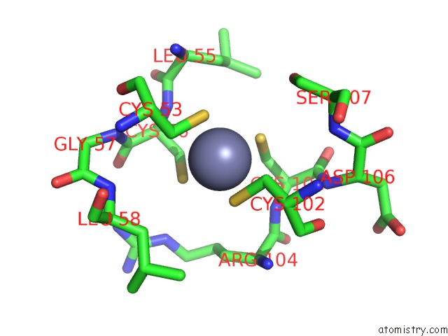
Mono view
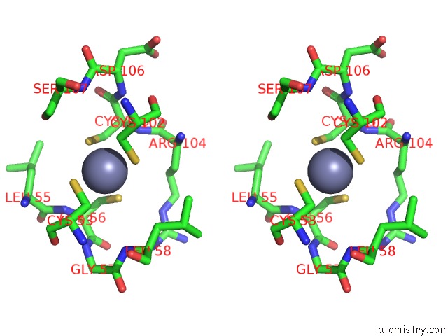
Stereo pair view
Mono view
Stereo pair view
A full contact list of Zinc with other atoms in the Zn binding
site number 1 of Structure of Msrb From Xanthomonas Campestris (Oxidized Form) within 5.0Å range:
|
Zinc binding site 2 out of 2 in 3hcj
Go back to
Zinc binding site 2 out
of 2 in the Structure of Msrb From Xanthomonas Campestris (Oxidized Form)
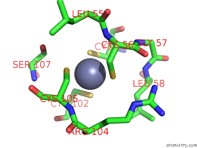
Mono view

Stereo pair view
Mono view
Stereo pair view
A full contact list of Zinc with other atoms in the Zn binding
site number 2 of Structure of Msrb From Xanthomonas Campestris (Oxidized Form) within 5.0Å range:
|
Reference:
F.M.Ranaivoson,
F.Neiers,
B.Kauffmann,
S.Boschi-Muller,
G.Branlant,
F.Favier.
Methionine Sulfoxide Reductase B Displays A High Level of Flexibility. J.Mol.Biol. 2009.
ISSN: ESSN 1089-8638
PubMed: 19733575
DOI: 10.1016/J.JMB.2009.08.073
Page generated: Wed Aug 20 10:03:07 2025
ISSN: ESSN 1089-8638
PubMed: 19733575
DOI: 10.1016/J.JMB.2009.08.073
Last articles
Zn in 3QLNZn in 3QMC
Zn in 3QMB
Zn in 3QLC
Zn in 3QLA
Zn in 3QL9
Zn in 3QJ5
Zn in 3QG6
Zn in 3QJX
Zn in 3QJ0