Zinc »
PDB 3h6t-3hk5 »
3h9b »
Zinc in PDB 3h9b: Structure of A Mutant Methionyl-Trna Synthetase with Modified Specificity Complexed with Azidonorleucine
Enzymatic activity of Structure of A Mutant Methionyl-Trna Synthetase with Modified Specificity Complexed with Azidonorleucine
All present enzymatic activity of Structure of A Mutant Methionyl-Trna Synthetase with Modified Specificity Complexed with Azidonorleucine:
6.1.1.10;
6.1.1.10;
Protein crystallography data
The structure of Structure of A Mutant Methionyl-Trna Synthetase with Modified Specificity Complexed with Azidonorleucine, PDB code: 3h9b
was solved by
E.Schmitt,
I.C.Tanrikulu,
T.H.Yoo,
M.Panvert,
D.A.Tirrell,
Y.Mechulam,
with X-Ray Crystallography technique. A brief refinement statistics is given in the table below:
| Resolution Low / High (Å) | 50.00 / 1.50 |
| Space group | P 1 21 1 |
| Cell size a, b, c (Å), α, β, γ (°) | 78.259, 45.377, 85.890, 90.00, 107.24, 90.00 |
| R / Rfree (%) | 16.5 / 18.8 |
Zinc Binding Sites:
The binding sites of Zinc atom in the Structure of A Mutant Methionyl-Trna Synthetase with Modified Specificity Complexed with Azidonorleucine
(pdb code 3h9b). This binding sites where shown within
5.0 Angstroms radius around Zinc atom.
In total only one binding site of Zinc was determined in the Structure of A Mutant Methionyl-Trna Synthetase with Modified Specificity Complexed with Azidonorleucine, PDB code: 3h9b:
In total only one binding site of Zinc was determined in the Structure of A Mutant Methionyl-Trna Synthetase with Modified Specificity Complexed with Azidonorleucine, PDB code: 3h9b:
Zinc binding site 1 out of 1 in 3h9b
Go back to
Zinc binding site 1 out
of 1 in the Structure of A Mutant Methionyl-Trna Synthetase with Modified Specificity Complexed with Azidonorleucine
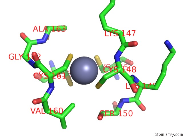
Mono view
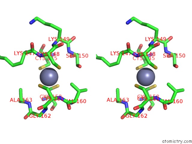
Stereo pair view
Mono view
Stereo pair view
A full contact list of Zinc with other atoms in the Zn binding
site number 1 of Structure of A Mutant Methionyl-Trna Synthetase with Modified Specificity Complexed with Azidonorleucine within 5.0Å range:
|
Reference:
E.Schmitt,
I.C.Tanrikulu,
T.H.Yoo,
M.Panvert,
D.A.Tirrell,
Y.Mechulam.
Switching From An Induced-Fit to A Lock-and-Key Mechanism in An Aminoacyl-Trna Synthetase with Modified Specificity. J.Mol.Biol. V. 394 843 2009.
ISSN: ISSN 0022-2836
PubMed: 19837083
DOI: 10.1016/J.JMB.2009.10.016
Page generated: Wed Aug 20 10:00:29 2025
ISSN: ISSN 0022-2836
PubMed: 19837083
DOI: 10.1016/J.JMB.2009.10.016
Last articles
Zn in 4C1HZn in 4C1D
Zn in 4C11
Zn in 4BZI
Zn in 4C1C
Zn in 4C09
Zn in 4BZS
Zn in 4BZ9
Zn in 4BZ8
Zn in 4BZR