Zinc in PDB 3g9o: Gr Dna-Binding Domain:Sgk 16BP Complex-9
Protein crystallography data
The structure of Gr Dna-Binding Domain:Sgk 16BP Complex-9, PDB code: 3g9o
was solved by
M.A.Pufall,
K.R.Yamamoto,
S.H.Meijsing,
with X-Ray Crystallography technique. A brief refinement statistics is given in the table below:
Resolution Low / High (Å)
|
34.35 /
1.65
|
Space group
|
C 1 2 1
|
Cell size a, b, c (Å), α, β, γ (°)
|
117.382,
38.470,
97.039,
90.00,
123.44,
90.00
|
R / Rfree (%)
|
17.7 /
20.3
|
Zinc Binding Sites:
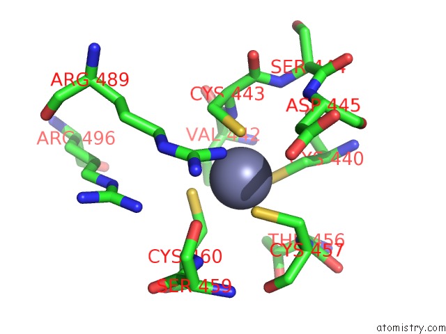
The binding sites of Zinc atom in the Gr Dna-Binding Domain:Sgk 16BP Complex-9
(pdb code 3g9o). This binding sites where shown within
5.0 Angstroms radius around Zinc atom.
In total 4 binding sites of Zinc where determined in the
Gr Dna-Binding Domain:Sgk 16BP Complex-9, PDB code: 3g9o:
Jump to Zinc binding site number:
1;
2;
3;
4;
Zinc binding site 1 out
of 4 in 3g9o
Go back to
Zinc Binding Sites List in 3g9o
Zinc binding site 1 out
of 4 in the Gr Dna-Binding Domain:Sgk 16BP Complex-9
Mono view
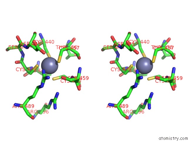
Stereo pair view
|
A full contact list of Zinc with other atoms in the Zn binding
site number 1 of Gr Dna-Binding Domain:Sgk 16BP Complex-9 within 5.0Å range:
probe
|
atom
|
residue
|
distance (Å)
|
B
|
Occ
|
B:Zn526
b:46.4
occ:1.00
|
SG
|
B:CYS443
|
2.3
|
48.0
|
1.0
|
SG
|
B:CYS457
|
2.3
|
47.0
|
1.0
|
SG
|
B:CYS440
|
2.4
|
47.0
|
1.0
|
SG
|
B:CYS460
|
2.4
|
40.7
|
1.0
|
CB
|
B:CYS460
|
3.1
|
47.5
|
1.0
|
CB
|
B:CYS440
|
3.2
|
63.6
|
1.0
|
CB
|
B:CYS443
|
3.3
|
48.0
|
1.0
|
CB
|
B:CYS457
|
3.5
|
52.6
|
1.0
|
N
|
B:CYS443
|
3.7
|
44.7
|
1.0
|
N
|
B:CYS457
|
3.9
|
54.9
|
1.0
|
CA
|
B:CYS443
|
4.0
|
50.3
|
1.0
|
N
|
B:CYS460
|
4.2
|
40.0
|
1.0
|
CA
|
B:CYS460
|
4.2
|
43.1
|
1.0
|
NH1
|
B:ARG489
|
4.3
|
53.5
|
1.0
|
CA
|
B:CYS457
|
4.3
|
52.6
|
1.0
|
CB
|
B:VAL442
|
4.5
|
51.5
|
1.0
|
CB
|
B:ASP445
|
4.5
|
65.0
|
1.0
|
OG
|
B:SER459
|
4.6
|
52.1
|
1.0
|
C
|
B:CYS443
|
4.7
|
54.8
|
1.0
|
CA
|
B:CYS440
|
4.7
|
60.1
|
1.0
|
C
|
B:VAL442
|
4.7
|
42.4
|
1.0
|
N
|
B:SER444
|
4.8
|
55.0
|
1.0
|
N
|
B:ASP445
|
4.8
|
60.0
|
1.0
|
C
|
B:CYS457
|
4.9
|
52.3
|
1.0
|
O
|
B:CYS457
|
4.9
|
49.5
|
1.0
|
CD
|
B:ARG489
|
5.0
|
45.4
|
1.0
|
NH2
|
B:ARG496
|
5.0
|
35.4
|
1.0
|
CA
|
B:VAL442
|
5.0
|
47.7
|
1.0
|
C
|
B:THR456
|
5.0
|
59.3
|
1.0
|
|
Zinc binding site 2 out
of 4 in 3g9o
Go back to
Zinc Binding Sites List in 3g9o
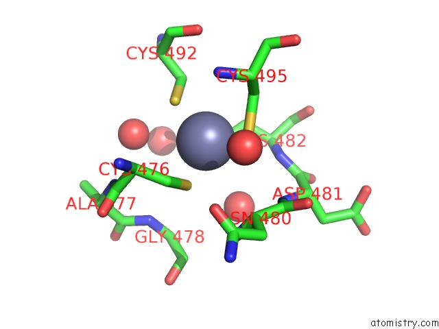
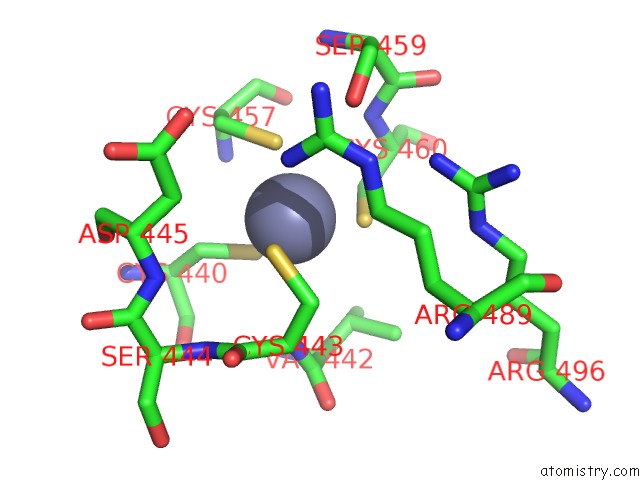
Zinc binding site 2 out
of 4 in the Gr Dna-Binding Domain:Sgk 16BP Complex-9
Mono view
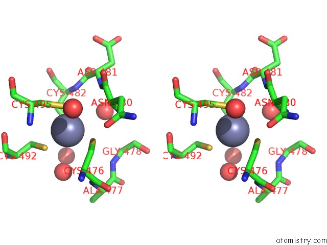
Stereo pair view
|
A full contact list of Zinc with other atoms in the Zn binding
site number 2 of Gr Dna-Binding Domain:Sgk 16BP Complex-9 within 5.0Å range:
probe
|
atom
|
residue
|
distance (Å)
|
B
|
Occ
|
B:Zn527
b:29.2
occ:1.00
|
SG
|
B:CYS476
|
2.3
|
26.3
|
1.0
|
SG
|
B:CYS495
|
2.3
|
28.6
|
1.0
|
SG
|
B:CYS482
|
2.3
|
32.0
|
1.0
|
SG
|
B:CYS492
|
2.4
|
27.9
|
1.0
|
CB
|
B:CYS482
|
3.2
|
34.7
|
1.0
|
CB
|
B:CYS476
|
3.3
|
23.8
|
1.0
|
CB
|
B:CYS495
|
3.3
|
27.2
|
1.0
|
CB
|
B:CYS492
|
3.4
|
25.1
|
1.0
|
O
|
B:HOH3
|
3.8
|
29.0
|
1.0
|
CA
|
B:CYS476
|
4.0
|
21.7
|
1.0
|
N
|
B:CYS482
|
4.0
|
31.1
|
1.0
|
O
|
B:HOH1
|
4.1
|
31.5
|
1.0
|
N
|
B:CYS495
|
4.2
|
26.7
|
1.0
|
CA
|
B:CYS482
|
4.2
|
29.9
|
1.0
|
CA
|
B:CYS495
|
4.3
|
29.6
|
1.0
|
N
|
B:ASP481
|
4.6
|
27.0
|
1.0
|
O
|
A:HOH32
|
4.6
|
40.3
|
1.0
|
CA
|
B:ASN480
|
4.6
|
29.4
|
1.0
|
O
|
B:HOH2
|
4.7
|
35.8
|
1.0
|
N
|
B:ALA477
|
4.7
|
26.7
|
1.0
|
N
|
B:ASN480
|
4.7
|
28.2
|
1.0
|
CA
|
B:CYS492
|
4.8
|
26.3
|
1.0
|
C
|
B:CYS476
|
4.8
|
23.0
|
1.0
|
N
|
B:GLY478
|
4.9
|
27.5
|
1.0
|
C
|
B:ASN480
|
4.9
|
32.1
|
1.0
|
|
Zinc binding site 3 out
of 4 in 3g9o
Go back to
Zinc Binding Sites List in 3g9o
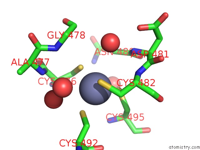
Zinc binding site 3 out
of 4 in the Gr Dna-Binding Domain:Sgk 16BP Complex-9
Mono view
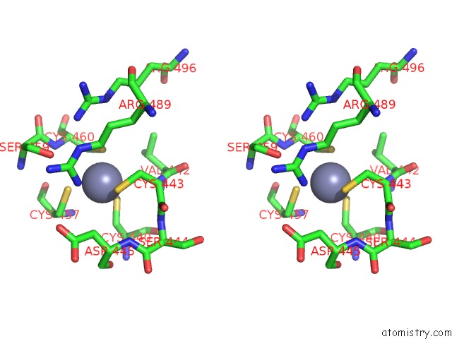
Stereo pair view
|
A full contact list of Zinc with other atoms in the Zn binding
site number 3 of Gr Dna-Binding Domain:Sgk 16BP Complex-9 within 5.0Å range:
probe
|
atom
|
residue
|
distance (Å)
|
B
|
Occ
|
A:Zn526
b:33.4
occ:1.00
|
SG
|
A:CYS460
|
2.3
|
32.4
|
1.0
|
SG
|
A:CYS443
|
2.3
|
33.8
|
1.0
|
SG
|
A:CYS440
|
2.4
|
30.6
|
1.0
|
SG
|
A:CYS457
|
2.4
|
33.0
|
1.0
|
CB
|
A:CYS460
|
3.2
|
31.7
|
1.0
|
CB
|
A:CYS440
|
3.3
|
36.5
|
1.0
|
CB
|
A:CYS443
|
3.3
|
33.1
|
1.0
|
CB
|
A:CYS457
|
3.6
|
33.3
|
1.0
|
N
|
A:CYS443
|
3.7
|
32.1
|
1.0
|
N
|
A:CYS457
|
3.9
|
32.6
|
1.0
|
CA
|
A:CYS443
|
4.0
|
34.2
|
1.0
|
N
|
A:CYS460
|
4.2
|
29.6
|
1.0
|
NH1
|
A:ARG489
|
4.2
|
37.6
|
1.0
|
CA
|
A:CYS460
|
4.3
|
30.1
|
1.0
|
CA
|
A:CYS457
|
4.3
|
33.5
|
1.0
|
OG
|
A:SER459
|
4.4
|
36.8
|
1.0
|
CB
|
A:VAL442
|
4.5
|
30.6
|
1.0
|
CB
|
A:ASP445
|
4.5
|
49.9
|
1.0
|
C
|
A:CYS443
|
4.6
|
42.7
|
1.0
|
C
|
A:VAL442
|
4.7
|
33.5
|
1.0
|
CA
|
A:CYS440
|
4.7
|
31.7
|
1.0
|
N
|
A:SER444
|
4.8
|
39.0
|
1.0
|
C
|
A:CYS457
|
4.9
|
36.1
|
1.0
|
N
|
A:ASP445
|
4.9
|
45.8
|
1.0
|
O
|
A:CYS457
|
4.9
|
32.5
|
1.0
|
NH2
|
A:ARG496
|
4.9
|
28.2
|
1.0
|
CD
|
A:ARG489
|
5.0
|
28.2
|
1.0
|
CA
|
A:VAL442
|
5.0
|
30.2
|
1.0
|
|
Zinc binding site 4 out
of 4 in 3g9o
Go back to
Zinc Binding Sites List in 3g9o
Zinc binding site 4 out
of 4 in the Gr Dna-Binding Domain:Sgk 16BP Complex-9
Mono view
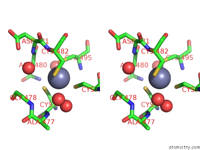
Stereo pair view
|
A full contact list of Zinc with other atoms in the Zn binding
site number 4 of Gr Dna-Binding Domain:Sgk 16BP Complex-9 within 5.0Å range:
probe
|
atom
|
residue
|
distance (Å)
|
B
|
Occ
|
A:Zn527
b:30.1
occ:1.00
|
SG
|
A:CYS482
|
2.3
|
31.0
|
1.0
|
SG
|
A:CYS476
|
2.3
|
28.9
|
1.0
|
SG
|
A:CYS495
|
2.4
|
28.2
|
1.0
|
SG
|
A:CYS492
|
2.4
|
28.0
|
1.0
|
CB
|
A:CYS482
|
3.2
|
30.3
|
1.0
|
CB
|
A:CYS476
|
3.3
|
31.2
|
1.0
|
CB
|
A:CYS492
|
3.3
|
27.4
|
1.0
|
CB
|
A:CYS495
|
3.5
|
27.2
|
1.0
|
O
|
A:HOH7
|
3.8
|
33.7
|
1.0
|
N
|
A:CYS482
|
4.0
|
33.3
|
1.0
|
CA
|
A:CYS476
|
4.0
|
28.8
|
1.0
|
N
|
A:CYS495
|
4.2
|
25.9
|
1.0
|
CA
|
A:CYS482
|
4.2
|
32.3
|
1.0
|
CA
|
A:ASN480
|
4.3
|
38.4
|
1.0
|
CA
|
A:CYS495
|
4.4
|
24.2
|
1.0
|
N
|
A:ASN480
|
4.5
|
35.5
|
1.0
|
O
|
A:HOH11
|
4.6
|
31.5
|
1.0
|
O
|
A:HOH21
|
4.6
|
45.7
|
1.0
|
N
|
A:ASP481
|
4.6
|
31.5
|
1.0
|
N
|
A:ALA477
|
4.7
|
32.3
|
1.0
|
O
|
A:HOH32
|
4.7
|
40.3
|
1.0
|
CA
|
A:CYS492
|
4.7
|
25.1
|
1.0
|
C
|
A:ASN480
|
4.7
|
33.6
|
1.0
|
N
|
A:GLY478
|
4.8
|
33.7
|
1.0
|
C
|
A:CYS476
|
4.8
|
32.1
|
1.0
|
|
Reference:
S.H.Meijsing,
M.A.Pufall,
A.Y.So,
D.L.Bates,
L.Chen,
K.R.Yamamoto.
Dna Binding Site Sequence Directs Glucocorticoid Receptor Structure and Activity. Science V. 324 407 2009.
ISSN: ISSN 0036-8075
PubMed: 19372434
DOI: 10.1126/SCIENCE.1164265
Page generated: Thu Oct 24 13:39:05 2024
|