Zinc »
PDB 3flf-3fuk »
3ftv »
Zinc in PDB 3ftv: Leukotriene A4 Hydrolase in Complex with Fragment N- (Pyridin-3-Ylmethyl)Aniline
Enzymatic activity of Leukotriene A4 Hydrolase in Complex with Fragment N- (Pyridin-3-Ylmethyl)Aniline
All present enzymatic activity of Leukotriene A4 Hydrolase in Complex with Fragment N- (Pyridin-3-Ylmethyl)Aniline:
3.3.2.6;
3.3.2.6;
Protein crystallography data
The structure of Leukotriene A4 Hydrolase in Complex with Fragment N- (Pyridin-3-Ylmethyl)Aniline, PDB code: 3ftv
was solved by
D.R.Davies,
with X-Ray Crystallography technique. A brief refinement statistics is given in the table below:
| Resolution Low / High (Å) | 50.00 / 1.70 |
| Space group | P 21 21 21 |
| Cell size a, b, c (Å), α, β, γ (°) | 78.124, 87.000, 99.328, 90.00, 90.00, 90.00 |
| R / Rfree (%) | 17.5 / 20.6 |
Other elements in 3ftv:
The structure of Leukotriene A4 Hydrolase in Complex with Fragment N- (Pyridin-3-Ylmethyl)Aniline also contains other interesting chemical elements:
| Ytterbium | (Yb) | 2 atoms |
Zinc Binding Sites:
The binding sites of Zinc atom in the Leukotriene A4 Hydrolase in Complex with Fragment N- (Pyridin-3-Ylmethyl)Aniline
(pdb code 3ftv). This binding sites where shown within
5.0 Angstroms radius around Zinc atom.
In total only one binding site of Zinc was determined in the Leukotriene A4 Hydrolase in Complex with Fragment N- (Pyridin-3-Ylmethyl)Aniline, PDB code: 3ftv:
In total only one binding site of Zinc was determined in the Leukotriene A4 Hydrolase in Complex with Fragment N- (Pyridin-3-Ylmethyl)Aniline, PDB code: 3ftv:
Zinc binding site 1 out of 1 in 3ftv
Go back to
Zinc binding site 1 out
of 1 in the Leukotriene A4 Hydrolase in Complex with Fragment N- (Pyridin-3-Ylmethyl)Aniline
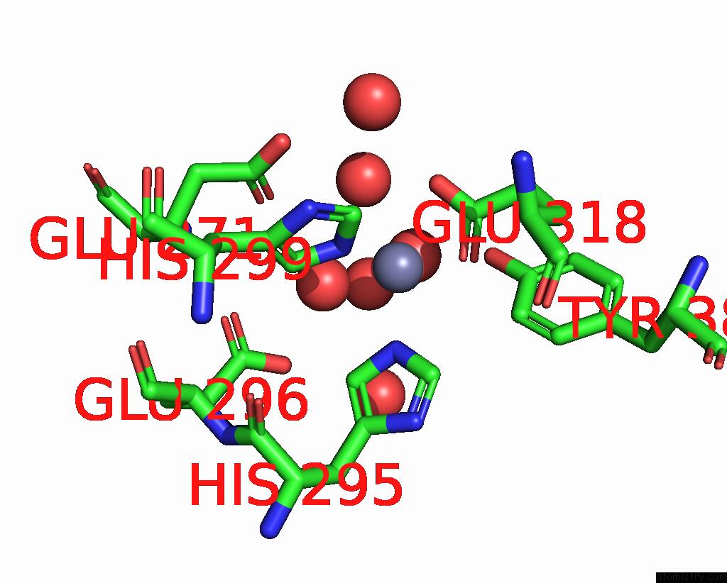
Mono view
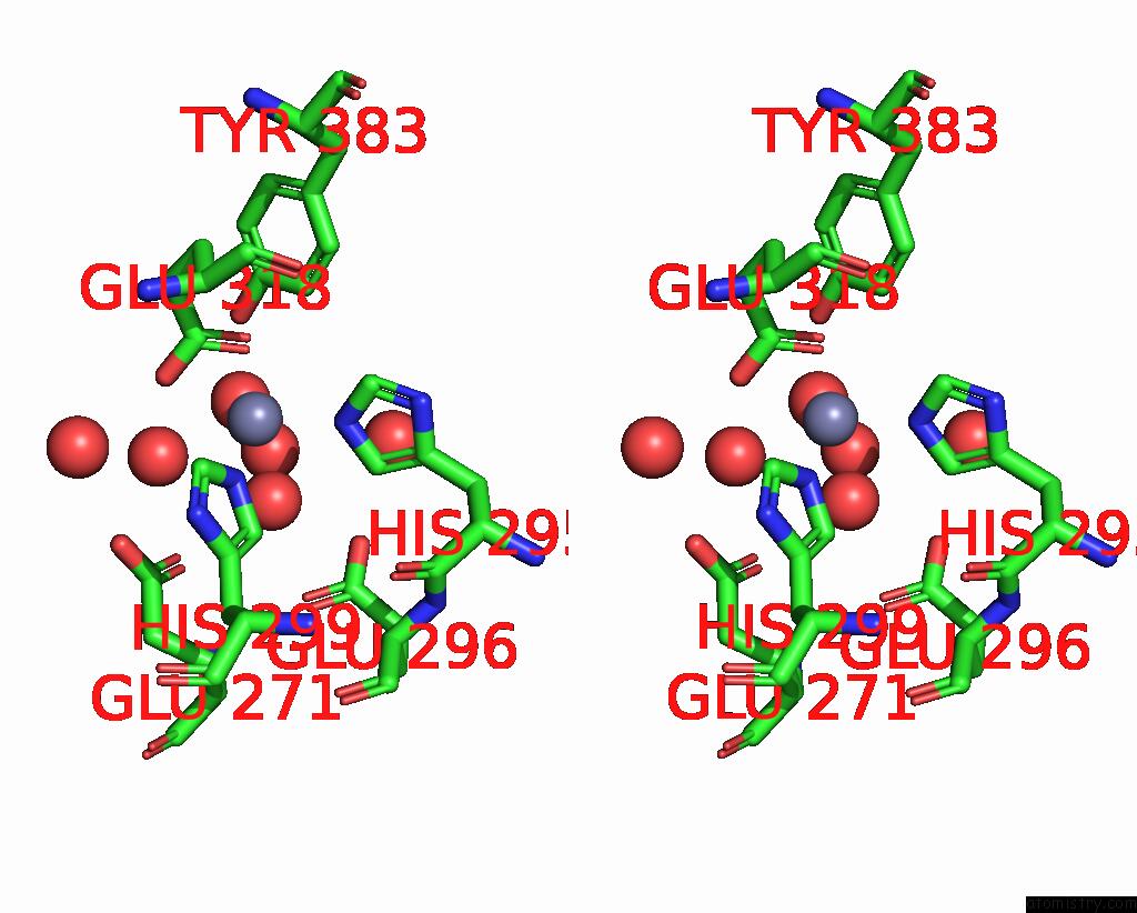
Stereo pair view
Mono view
Stereo pair view
A full contact list of Zinc with other atoms in the Zn binding
site number 1 of Leukotriene A4 Hydrolase in Complex with Fragment N- (Pyridin-3-Ylmethyl)Aniline within 5.0Å range:
|
Reference:
D.R.Davies,
B.Mamat,
O.T.Magnusson,
J.Christensen,
M.H.Haraldsson,
R.Mishra,
B.Pease,
E.Hansen,
J.Singh,
D.Zembower,
H.Kim,
A.S.Kiselyov,
A.B.Burgin,
M.E.Gurney,
L.J.Stewart.
Discovery of Leukotriene A4 Hydrolase Inhibitors Using Metabolomics Biased Fragment Crystallography. J.Med.Chem. V. 52 4694 2009.
ISSN: ISSN 0022-2623
PubMed: 19618939
DOI: 10.1021/JM900259H
Page generated: Thu Oct 24 13:24:20 2024
ISSN: ISSN 0022-2623
PubMed: 19618939
DOI: 10.1021/JM900259H
Last articles
Zn in 9MJ5Zn in 9HNW
Zn in 9G0L
Zn in 9FNE
Zn in 9DZN
Zn in 9E0I
Zn in 9D32
Zn in 9DAK
Zn in 8ZXC
Zn in 8ZUF