Zinc »
PDB 3ekz-3eyv »
3eqo »
Zinc in PDB 3eqo: Crystal Structure of Beta-1,3-Glucanase From Phanerochaete Chrysosporium (LAM55A) Gluconolactone Complex
Enzymatic activity of Crystal Structure of Beta-1,3-Glucanase From Phanerochaete Chrysosporium (LAM55A) Gluconolactone Complex
All present enzymatic activity of Crystal Structure of Beta-1,3-Glucanase From Phanerochaete Chrysosporium (LAM55A) Gluconolactone Complex:
3.2.1.58;
3.2.1.58;
Protein crystallography data
The structure of Crystal Structure of Beta-1,3-Glucanase From Phanerochaete Chrysosporium (LAM55A) Gluconolactone Complex, PDB code: 3eqo
was solved by
T.Ishida,
S.Fushinobu,
R.Kawai,
M.Kitaoka,
K.Igarashi,
M.Samejima,
with X-Ray Crystallography technique. A brief refinement statistics is given in the table below:
| Resolution Low / High (Å) | 31.50 / 2.25 |
| Space group | P 1 |
| Cell size a, b, c (Å), α, β, γ (°) | 66.534, 67.146, 109.778, 93.94, 106.76, 97.08 |
| R / Rfree (%) | 14.2 / 20 |
Zinc Binding Sites:
The binding sites of Zinc atom in the Crystal Structure of Beta-1,3-Glucanase From Phanerochaete Chrysosporium (LAM55A) Gluconolactone Complex
(pdb code 3eqo). This binding sites where shown within
5.0 Angstroms radius around Zinc atom.
In total 2 binding sites of Zinc where determined in the Crystal Structure of Beta-1,3-Glucanase From Phanerochaete Chrysosporium (LAM55A) Gluconolactone Complex, PDB code: 3eqo:
Jump to Zinc binding site number: 1; 2;
In total 2 binding sites of Zinc where determined in the Crystal Structure of Beta-1,3-Glucanase From Phanerochaete Chrysosporium (LAM55A) Gluconolactone Complex, PDB code: 3eqo:
Jump to Zinc binding site number: 1; 2;
Zinc binding site 1 out of 2 in 3eqo
Go back to
Zinc binding site 1 out
of 2 in the Crystal Structure of Beta-1,3-Glucanase From Phanerochaete Chrysosporium (LAM55A) Gluconolactone Complex
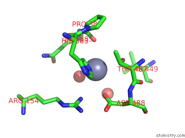
Mono view
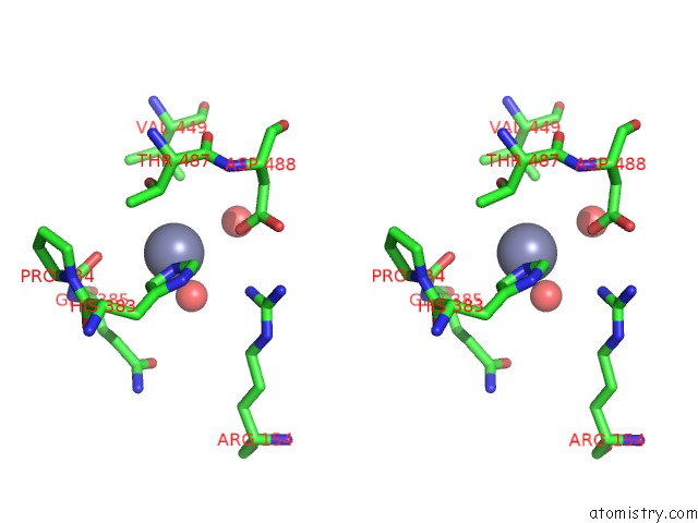
Stereo pair view
Mono view
Stereo pair view
A full contact list of Zinc with other atoms in the Zn binding
site number 1 of Crystal Structure of Beta-1,3-Glucanase From Phanerochaete Chrysosporium (LAM55A) Gluconolactone Complex within 5.0Å range:
|
Zinc binding site 2 out of 2 in 3eqo
Go back to
Zinc binding site 2 out
of 2 in the Crystal Structure of Beta-1,3-Glucanase From Phanerochaete Chrysosporium (LAM55A) Gluconolactone Complex
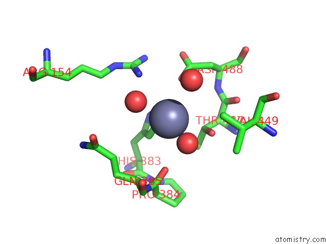
Mono view
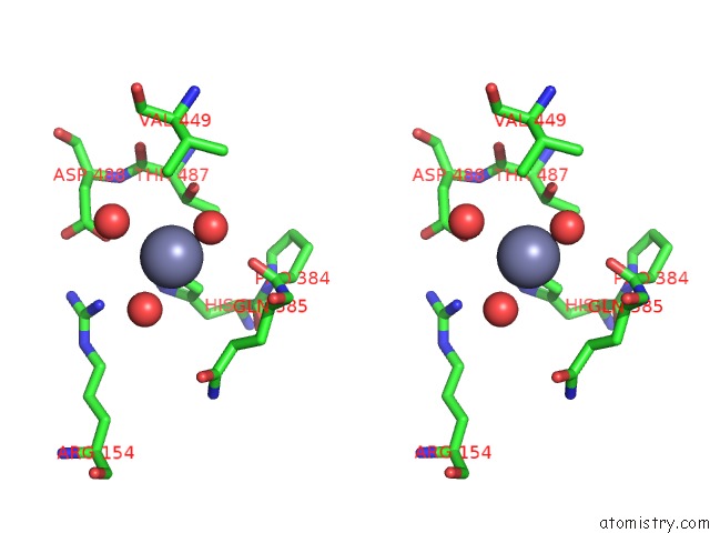
Stereo pair view
Mono view
Stereo pair view
A full contact list of Zinc with other atoms in the Zn binding
site number 2 of Crystal Structure of Beta-1,3-Glucanase From Phanerochaete Chrysosporium (LAM55A) Gluconolactone Complex within 5.0Å range:
|
Reference:
T.Ishida,
S.Fushinobu,
R.Kawai,
M.Kitaoka,
K.Igarashi,
M.Samejima.
Crystal Structure of Glycoside Hydrolase Family 55 Beta -1,3-Glucanase From the Basidiomycete Phanerochaete Chrysosporium J.Biol.Chem. V. 284 10100 2009.
ISSN: ISSN 0021-9258
PubMed: 19193645
DOI: 10.1074/JBC.M808122200
Page generated: Thu Oct 24 12:54:10 2024
ISSN: ISSN 0021-9258
PubMed: 19193645
DOI: 10.1074/JBC.M808122200
Last articles
Zn in 9MJ5Zn in 9HNW
Zn in 9G0L
Zn in 9FNE
Zn in 9DZN
Zn in 9E0I
Zn in 9D32
Zn in 9DAK
Zn in 8ZXC
Zn in 8ZUF