Zinc »
PDB 3ekz-3eyv »
3eo3 »
Zinc in PDB 3eo3: Crystal Structure of the N-Acetylmannosamine Kinase Domain of Human Gne Protein
Enzymatic activity of Crystal Structure of the N-Acetylmannosamine Kinase Domain of Human Gne Protein
All present enzymatic activity of Crystal Structure of the N-Acetylmannosamine Kinase Domain of Human Gne Protein:
2.7.1.60; 5.1.3.14;
2.7.1.60; 5.1.3.14;
Protein crystallography data
The structure of Crystal Structure of the N-Acetylmannosamine Kinase Domain of Human Gne Protein, PDB code: 3eo3
was solved by
L.Nedyalkova,
Y.Tong,
W.M.Rabeh,
B.Hong,
W.Tempel,
F.Mackenzie,
C.H.Arrowsmith,
A.M.Edwards,
C.Bountra,
J.Weigelt,
A.Bochkarev,
H.Park,
Structural Genomics Consortium (Sgc),
with X-Ray Crystallography technique. A brief refinement statistics is given in the table below:
| Resolution Low / High (Å) | 30.00 / 2.84 |
| Space group | P 31 2 1 |
| Cell size a, b, c (Å), α, β, γ (°) | 127.946, 127.946, 127.247, 90.00, 90.00, 120.00 |
| R / Rfree (%) | 20.5 / 24.5 |
Zinc Binding Sites:
The binding sites of Zinc atom in the Crystal Structure of the N-Acetylmannosamine Kinase Domain of Human Gne Protein
(pdb code 3eo3). This binding sites where shown within
5.0 Angstroms radius around Zinc atom.
In total 3 binding sites of Zinc where determined in the Crystal Structure of the N-Acetylmannosamine Kinase Domain of Human Gne Protein, PDB code: 3eo3:
Jump to Zinc binding site number: 1; 2; 3;
In total 3 binding sites of Zinc where determined in the Crystal Structure of the N-Acetylmannosamine Kinase Domain of Human Gne Protein, PDB code: 3eo3:
Jump to Zinc binding site number: 1; 2; 3;
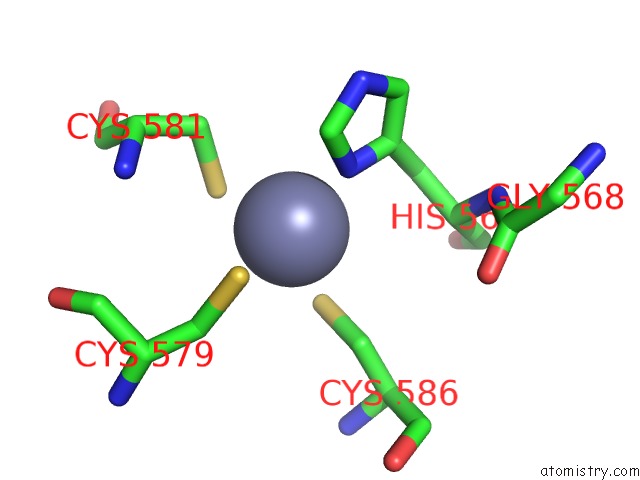
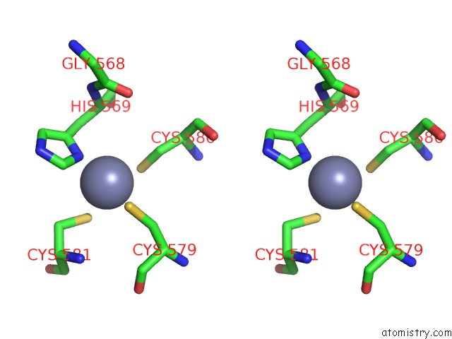
Zinc binding site 1 out of 3 in 3eo3
Go back to
Zinc binding site 1 out
of 3 in the Crystal Structure of the N-Acetylmannosamine Kinase Domain of Human Gne Protein
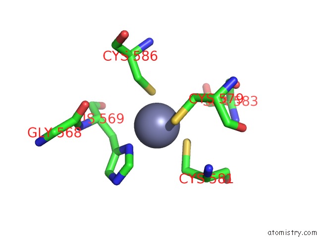
Mono view
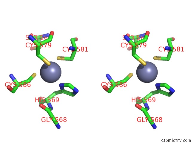
Stereo pair view
Mono view
Stereo pair view
A full contact list of Zinc with other atoms in the Zn binding
site number 1 of Crystal Structure of the N-Acetylmannosamine Kinase Domain of Human Gne Protein within 5.0Å range:
|
Zinc binding site 2 out of 3 in 3eo3
Go back to
Zinc binding site 2 out
of 3 in the Crystal Structure of the N-Acetylmannosamine Kinase Domain of Human Gne Protein
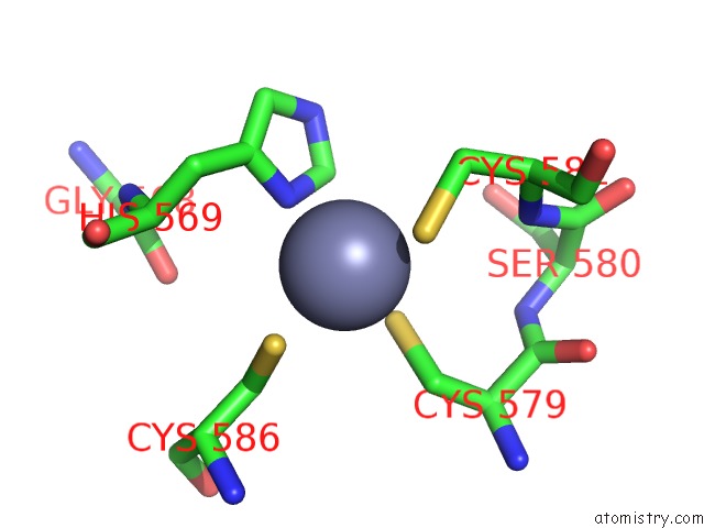
Mono view
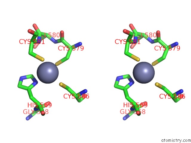
Stereo pair view
Mono view
Stereo pair view
A full contact list of Zinc with other atoms in the Zn binding
site number 2 of Crystal Structure of the N-Acetylmannosamine Kinase Domain of Human Gne Protein within 5.0Å range:
|
Zinc binding site 3 out of 3 in 3eo3
Go back to
Zinc binding site 3 out
of 3 in the Crystal Structure of the N-Acetylmannosamine Kinase Domain of Human Gne Protein
Mono view
Stereo pair view
Mono view
Stereo pair view
A full contact list of Zinc with other atoms in the Zn binding
site number 3 of Crystal Structure of the N-Acetylmannosamine Kinase Domain of Human Gne Protein within 5.0Å range:
|
Reference:
Y.Tong,
W.Tempel,
L.Nedyalkova,
F.Mackenzie,
H.W.Park.
Crystal Structure of the N-Acetylmannosamine Kinase Domain of Gne. Plos One V. 4 E7165 2009.
ISSN: ESSN 1932-6203
PubMed: 19841673
DOI: 10.1371/JOURNAL.PONE.0007165
Page generated: Thu Oct 24 12:52:12 2024
ISSN: ESSN 1932-6203
PubMed: 19841673
DOI: 10.1371/JOURNAL.PONE.0007165
Last articles
Zn in 9MJ5Zn in 9HNW
Zn in 9G0L
Zn in 9FNE
Zn in 9DZN
Zn in 9E0I
Zn in 9D32
Zn in 9DAK
Zn in 8ZXC
Zn in 8ZUF