Zinc »
PDB 3cjp-3czs »
3cqj »
Zinc in PDB 3cqj: Crystal Structure of L-Xylulose-5-Phosphate 3-Epimerase Ulae (Form B) Complex with ZN2+
Enzymatic activity of Crystal Structure of L-Xylulose-5-Phosphate 3-Epimerase Ulae (Form B) Complex with ZN2+
All present enzymatic activity of Crystal Structure of L-Xylulose-5-Phosphate 3-Epimerase Ulae (Form B) Complex with ZN2+:
5.1.3.22;
5.1.3.22;
Protein crystallography data
The structure of Crystal Structure of L-Xylulose-5-Phosphate 3-Epimerase Ulae (Form B) Complex with ZN2+, PDB code: 3cqj
was solved by
R.Shi,
A.Matte,
M.Cygler,
Montreal-Kingston Bacterial Structuralgenomics Initiative (Bsgi),
with X-Ray Crystallography technique. A brief refinement statistics is given in the table below:
| Resolution Low / High (Å) | 50.00 / 2.04 |
| Space group | C 2 2 21 |
| Cell size a, b, c (Å), α, β, γ (°) | 104.208, 132.596, 81.798, 90.00, 90.00, 90.00 |
| R / Rfree (%) | 18.3 / 21.3 |
Zinc Binding Sites:
The binding sites of Zinc atom in the Crystal Structure of L-Xylulose-5-Phosphate 3-Epimerase Ulae (Form B) Complex with ZN2+
(pdb code 3cqj). This binding sites where shown within
5.0 Angstroms radius around Zinc atom.
In total 4 binding sites of Zinc where determined in the Crystal Structure of L-Xylulose-5-Phosphate 3-Epimerase Ulae (Form B) Complex with ZN2+, PDB code: 3cqj:
Jump to Zinc binding site number: 1; 2; 3; 4;
In total 4 binding sites of Zinc where determined in the Crystal Structure of L-Xylulose-5-Phosphate 3-Epimerase Ulae (Form B) Complex with ZN2+, PDB code: 3cqj:
Jump to Zinc binding site number: 1; 2; 3; 4;
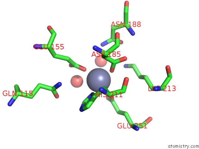
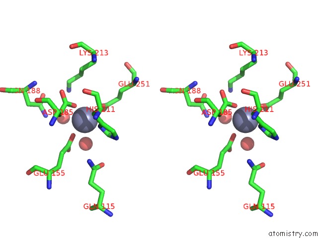
Zinc binding site 1 out of 4 in 3cqj
Go back to
Zinc binding site 1 out
of 4 in the Crystal Structure of L-Xylulose-5-Phosphate 3-Epimerase Ulae (Form B) Complex with ZN2+
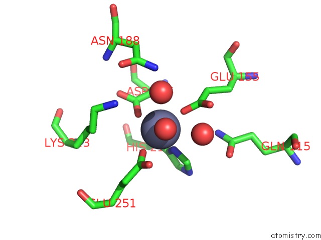
Mono view
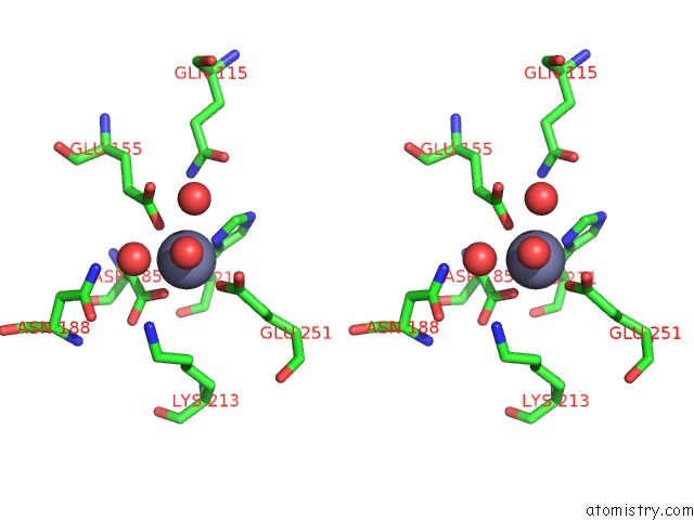
Stereo pair view
Mono view
Stereo pair view
A full contact list of Zinc with other atoms in the Zn binding
site number 1 of Crystal Structure of L-Xylulose-5-Phosphate 3-Epimerase Ulae (Form B) Complex with ZN2+ within 5.0Å range:
|
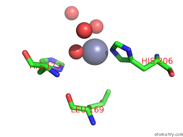
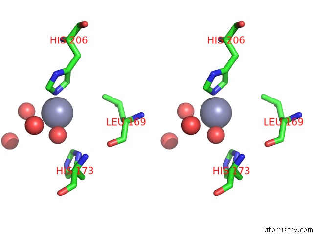
Zinc binding site 2 out of 4 in 3cqj
Go back to
Zinc binding site 2 out
of 4 in the Crystal Structure of L-Xylulose-5-Phosphate 3-Epimerase Ulae (Form B) Complex with ZN2+
Mono view
Stereo pair view
Mono view
Stereo pair view
A full contact list of Zinc with other atoms in the Zn binding
site number 2 of Crystal Structure of L-Xylulose-5-Phosphate 3-Epimerase Ulae (Form B) Complex with ZN2+ within 5.0Å range:
|
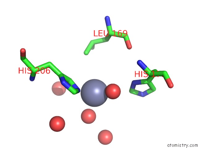
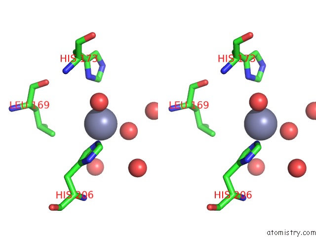
Zinc binding site 3 out of 4 in 3cqj
Go back to
Zinc binding site 3 out
of 4 in the Crystal Structure of L-Xylulose-5-Phosphate 3-Epimerase Ulae (Form B) Complex with ZN2+
Mono view
Stereo pair view
Mono view
Stereo pair view
A full contact list of Zinc with other atoms in the Zn binding
site number 3 of Crystal Structure of L-Xylulose-5-Phosphate 3-Epimerase Ulae (Form B) Complex with ZN2+ within 5.0Å range:
|
Zinc binding site 4 out of 4 in 3cqj
Go back to
Zinc binding site 4 out
of 4 in the Crystal Structure of L-Xylulose-5-Phosphate 3-Epimerase Ulae (Form B) Complex with ZN2+
Mono view
Stereo pair view
Mono view
Stereo pair view
A full contact list of Zinc with other atoms in the Zn binding
site number 4 of Crystal Structure of L-Xylulose-5-Phosphate 3-Epimerase Ulae (Form B) Complex with ZN2+ within 5.0Å range:
|
Reference:
R.Shi,
M.Pineda,
E.Ajamian,
Q.Cui,
A.Matte,
M.Cygler.
Structure of L-Xylulose-5-Phosphate 3-Epimerase (Ulae) From the Anaerobic L-Ascorbate Utilization Pathway of Escherichia Coli: Identification of A Novel Phosphate Binding Motif Within A Tim Barrel Fold. J.Bacteriol. V. 190 8137 2008.
ISSN: ISSN 0021-9193
PubMed: 18849419
DOI: 10.1128/JB.01049-08
Page generated: Thu Oct 24 11:54:45 2024
ISSN: ISSN 0021-9193
PubMed: 18849419
DOI: 10.1128/JB.01049-08
Last articles
Zn in 9MJ5Zn in 9HNW
Zn in 9G0L
Zn in 9FNE
Zn in 9DZN
Zn in 9E0I
Zn in 9D32
Zn in 9DAK
Zn in 8ZXC
Zn in 8ZUF