Zinc »
PDB 3cjp-3czs »
3cng »
Zinc in PDB 3cng: Crystal Structure of Nudix Hydrolase From Nitrosomonas Europaea
Protein crystallography data
The structure of Crystal Structure of Nudix Hydrolase From Nitrosomonas Europaea, PDB code: 3cng
was solved by
J.Osipiuk,
X.Xu,
H.Zheng,
A.Savchenko,
A.M.Edwards,
A.Joachimiak,
Midwestcenter For Structural Genomics (Mcsg),
with X-Ray Crystallography technique. A brief refinement statistics is given in the table below:
| Resolution Low / High (Å) | 37.20 / 2.00 |
| Space group | C 1 2 1 |
| Cell size a, b, c (Å), α, β, γ (°) | 174.781, 49.506, 110.190, 90.00, 106.28, 90.00 |
| R / Rfree (%) | 18.1 / 23.3 |
Other elements in 3cng:
The structure of Crystal Structure of Nudix Hydrolase From Nitrosomonas Europaea also contains other interesting chemical elements:
| Chlorine | (Cl) | 7 atoms |
| Sodium | (Na) | 2 atoms |
Zinc Binding Sites:
The binding sites of Zinc atom in the Crystal Structure of Nudix Hydrolase From Nitrosomonas Europaea
(pdb code 3cng). This binding sites where shown within
5.0 Angstroms radius around Zinc atom.
In total 4 binding sites of Zinc where determined in the Crystal Structure of Nudix Hydrolase From Nitrosomonas Europaea, PDB code: 3cng:
Jump to Zinc binding site number: 1; 2; 3; 4;
In total 4 binding sites of Zinc where determined in the Crystal Structure of Nudix Hydrolase From Nitrosomonas Europaea, PDB code: 3cng:
Jump to Zinc binding site number: 1; 2; 3; 4;
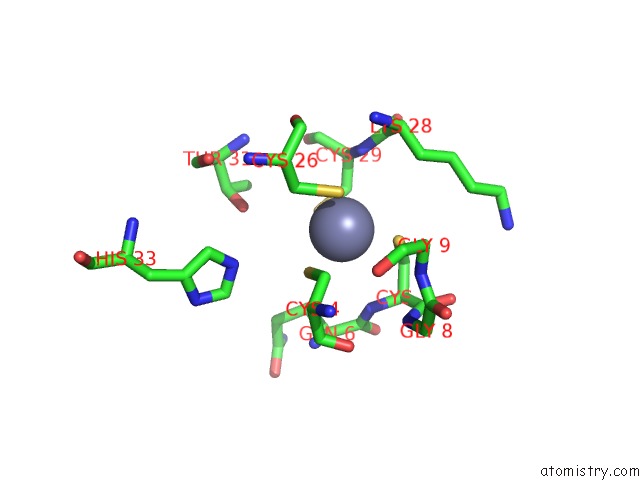
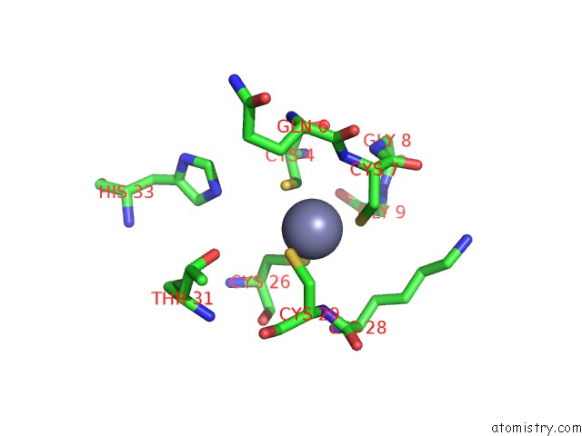
Zinc binding site 1 out of 4 in 3cng
Go back to
Zinc binding site 1 out
of 4 in the Crystal Structure of Nudix Hydrolase From Nitrosomonas Europaea
Mono view
Stereo pair view
Mono view
Stereo pair view
A full contact list of Zinc with other atoms in the Zn binding
site number 1 of Crystal Structure of Nudix Hydrolase From Nitrosomonas Europaea within 5.0Å range:
|
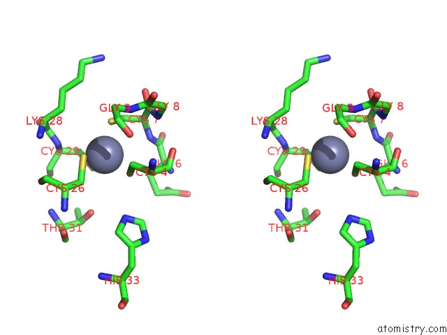
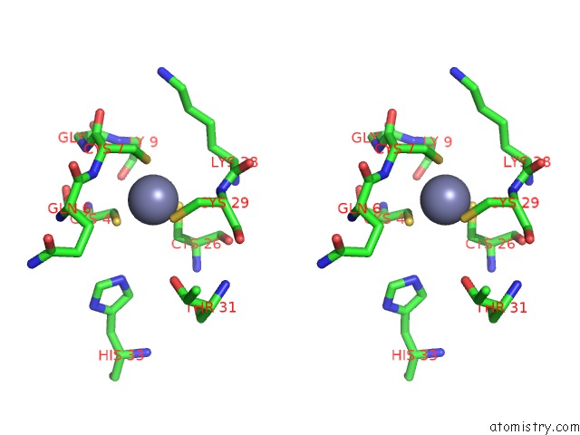
Zinc binding site 2 out of 4 in 3cng
Go back to
Zinc binding site 2 out
of 4 in the Crystal Structure of Nudix Hydrolase From Nitrosomonas Europaea
Mono view
Stereo pair view
Mono view
Stereo pair view
A full contact list of Zinc with other atoms in the Zn binding
site number 2 of Crystal Structure of Nudix Hydrolase From Nitrosomonas Europaea within 5.0Å range:
|
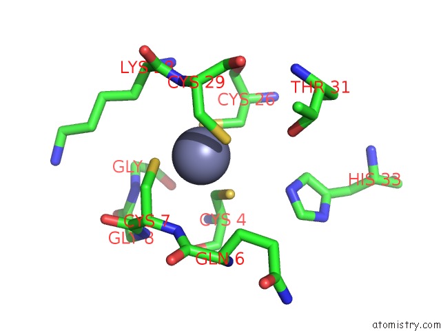
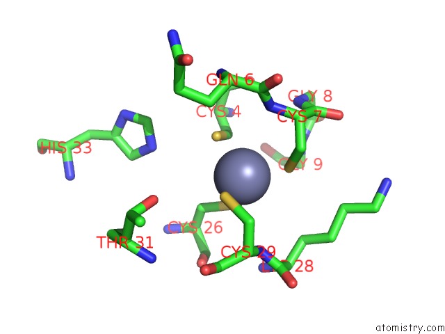
Zinc binding site 3 out of 4 in 3cng
Go back to
Zinc binding site 3 out
of 4 in the Crystal Structure of Nudix Hydrolase From Nitrosomonas Europaea
Mono view
Stereo pair view
Mono view
Stereo pair view
A full contact list of Zinc with other atoms in the Zn binding
site number 3 of Crystal Structure of Nudix Hydrolase From Nitrosomonas Europaea within 5.0Å range:
|
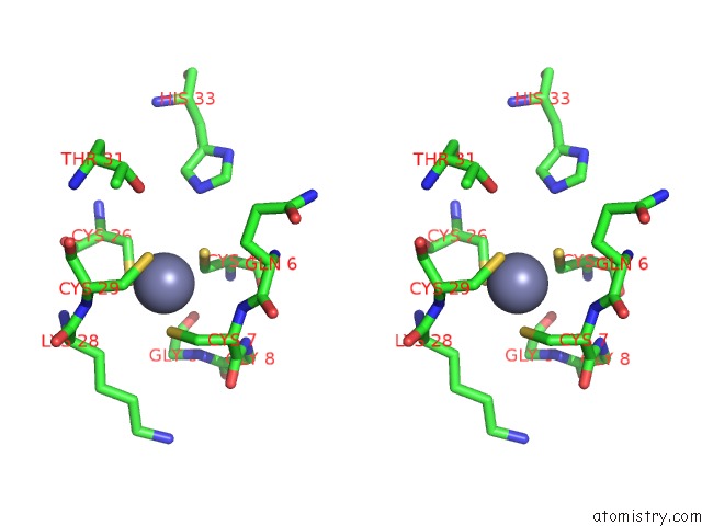
Zinc binding site 4 out of 4 in 3cng
Go back to
Zinc binding site 4 out
of 4 in the Crystal Structure of Nudix Hydrolase From Nitrosomonas Europaea
Mono view
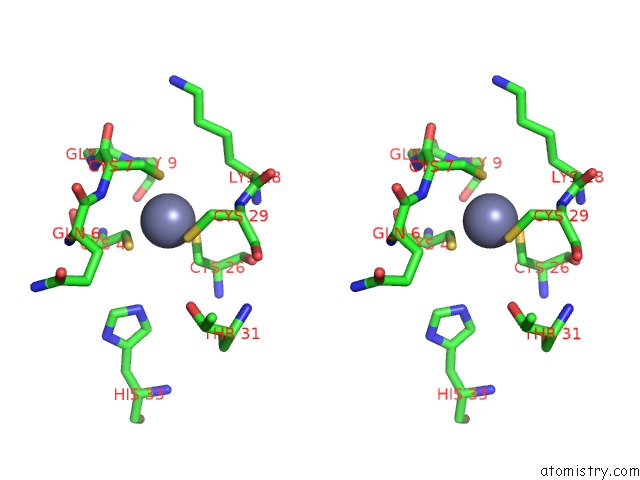
Stereo pair view
Mono view
Stereo pair view
A full contact list of Zinc with other atoms in the Zn binding
site number 4 of Crystal Structure of Nudix Hydrolase From Nitrosomonas Europaea within 5.0Å range:
|
Reference:
J.Osipiuk,
X.Xu,
H.Zheng,
A.Savchenko,
A.M.Edwards,
A.Joachimiak.
X-Ray Crystal Structure of Nudix Hydrolase From Nitrosomonas Europaea. To Be Published.
Page generated: Thu Oct 24 11:52:39 2024
Last articles
Al in 8SHAAl in 8SGL
Al in 8SH9
Al in 8SGC
Al in 8SG9
Al in 8SFF
Al in 8SG8
Al in 8R1A
Al in 8Q75
Al in 8OIE