Zinc »
PDB 3c52-3cjj »
3c7p »
Zinc in PDB 3c7p: Crystal Structure of Human Carbonic Anhydrase II in Complex with STX237
Enzymatic activity of Crystal Structure of Human Carbonic Anhydrase II in Complex with STX237
All present enzymatic activity of Crystal Structure of Human Carbonic Anhydrase II in Complex with STX237:
4.2.1.1;
4.2.1.1;
Protein crystallography data
The structure of Crystal Structure of Human Carbonic Anhydrase II in Complex with STX237, PDB code: 3c7p
was solved by
A.Di Fiore,
G.De Simone,
with X-Ray Crystallography technique. A brief refinement statistics is given in the table below:
| Resolution Low / High (Å) | 20.00 / 1.70 |
| Space group | P 1 21 1 |
| Cell size a, b, c (Å), α, β, γ (°) | 42.050, 41.440, 71.670, 90.00, 104.35, 90.00 |
| R / Rfree (%) | 18.1 / 20.2 |
Other elements in 3c7p:
The structure of Crystal Structure of Human Carbonic Anhydrase II in Complex with STX237 also contains other interesting chemical elements:
| Mercury | (Hg) | 1 atom |
| Chlorine | (Cl) | 1 atom |
Zinc Binding Sites:
The binding sites of Zinc atom in the Crystal Structure of Human Carbonic Anhydrase II in Complex with STX237
(pdb code 3c7p). This binding sites where shown within
5.0 Angstroms radius around Zinc atom.
In total only one binding site of Zinc was determined in the Crystal Structure of Human Carbonic Anhydrase II in Complex with STX237, PDB code: 3c7p:
In total only one binding site of Zinc was determined in the Crystal Structure of Human Carbonic Anhydrase II in Complex with STX237, PDB code: 3c7p:
Zinc binding site 1 out of 1 in 3c7p
Go back to
Zinc binding site 1 out
of 1 in the Crystal Structure of Human Carbonic Anhydrase II in Complex with STX237
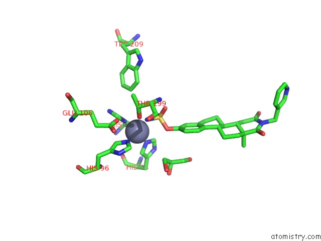
Mono view
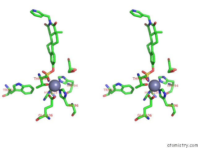
Stereo pair view
Mono view
Stereo pair view
A full contact list of Zinc with other atoms in the Zn binding
site number 1 of Crystal Structure of Human Carbonic Anhydrase II in Complex with STX237 within 5.0Å range:
|
Reference:
L.W.L.Woo,
D.S.Fischer,
C.M.Sharland,
M.Trusselle,
P.A.Foster,
S.K.Chander,
A.Di Fiore,
C.T.Supuran,
G.De Simone,
A.Purohit,
M.J.Reed,
B.V.L.Potter.
Anticancer Steroid Sulfatase Inhibitors: Synthesis of A Potent Fluorinated Second-Generation Agent, in Vitro and in Vivo Activities, Molecular Modeling, and Protein Crystallography Mol.Cancer Ther. V. 7 2435 2008.
ISSN: ISSN 1535-7163
PubMed: 18723489
DOI: 10.1158/1535-7163.MCT-08-0195
Page generated: Thu Oct 24 11:45:08 2024
ISSN: ISSN 1535-7163
PubMed: 18723489
DOI: 10.1158/1535-7163.MCT-08-0195
Last articles
Zn in 9MJ5Zn in 9HNW
Zn in 9G0L
Zn in 9FNE
Zn in 9DZN
Zn in 9E0I
Zn in 9D32
Zn in 9DAK
Zn in 8ZXC
Zn in 8ZUF