Zinc »
PDB 3bof-3c52 »
3bta »
Zinc in PDB 3bta: Crystal Structure of Botulinum Neurotoxin Serotype A
Enzymatic activity of Crystal Structure of Botulinum Neurotoxin Serotype A
All present enzymatic activity of Crystal Structure of Botulinum Neurotoxin Serotype A:
3.4.24.69;
3.4.24.69;
Protein crystallography data
The structure of Crystal Structure of Botulinum Neurotoxin Serotype A, PDB code: 3bta
was solved by
R.C.Stevens,
D.B.Lacy,
with X-Ray Crystallography technique. A brief refinement statistics is given in the table below:
| Resolution Low / High (Å) | 20.00 / 3.20 |
| Space group | P 31 2 1 |
| Cell size a, b, c (Å), α, β, γ (°) | 170.120, 170.120, 161.000, 90.00, 90.00, 120.00 |
| R / Rfree (%) | 22 / 28 |
Zinc Binding Sites:
The binding sites of Zinc atom in the Crystal Structure of Botulinum Neurotoxin Serotype A
(pdb code 3bta). This binding sites where shown within
5.0 Angstroms radius around Zinc atom.
In total only one binding site of Zinc was determined in the Crystal Structure of Botulinum Neurotoxin Serotype A, PDB code: 3bta:
In total only one binding site of Zinc was determined in the Crystal Structure of Botulinum Neurotoxin Serotype A, PDB code: 3bta:
Zinc binding site 1 out of 1 in 3bta
Go back to
Zinc binding site 1 out
of 1 in the Crystal Structure of Botulinum Neurotoxin Serotype A
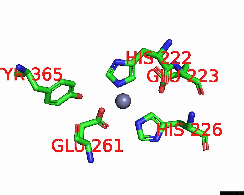
Mono view
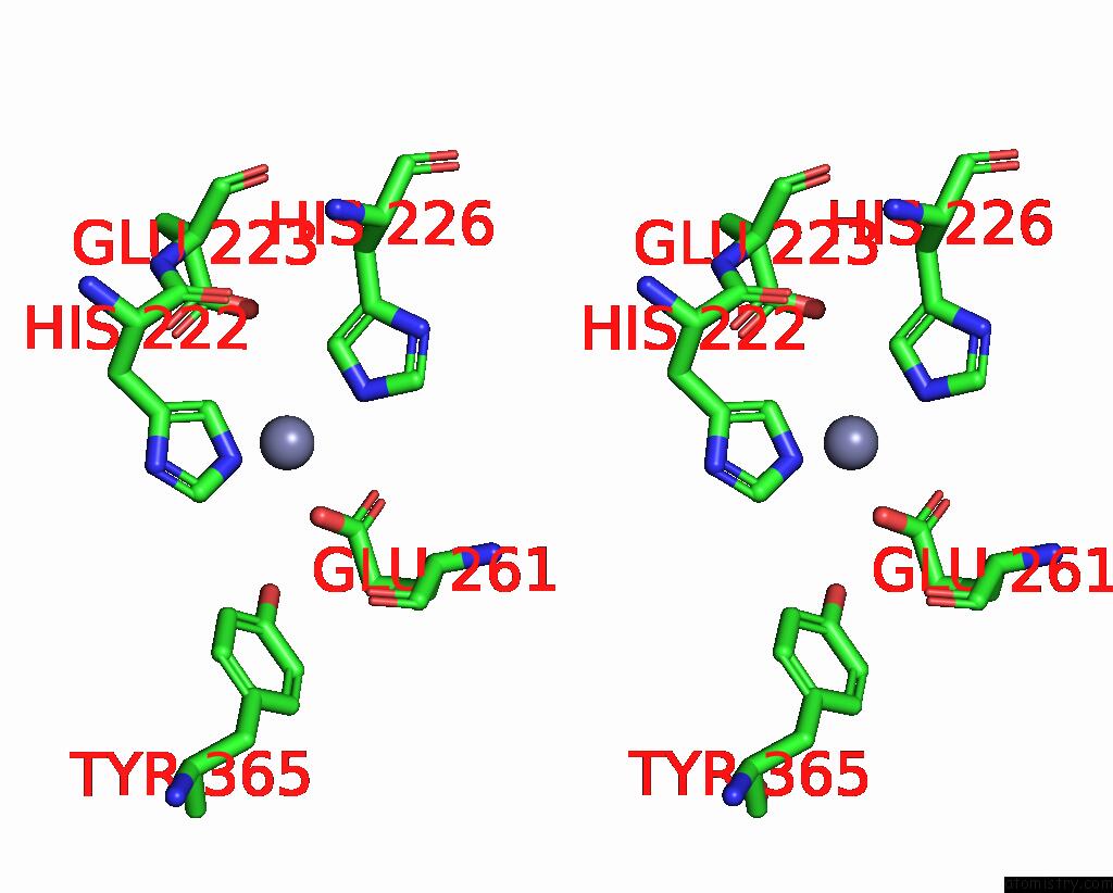
Stereo pair view
Mono view
Stereo pair view
A full contact list of Zinc with other atoms in the Zn binding
site number 1 of Crystal Structure of Botulinum Neurotoxin Serotype A within 5.0Å range:
|
Reference:
D.B.Lacy,
W.Tepp,
A.C.Cohen,
B.R.Dasgupta,
R.C.Stevens.
Crystal Structure of Botulinum Neurotoxin Type A and Implications For Toxicity. Nat.Struct.Biol. V. 5 898 1998.
ISSN: ISSN 1072-8368
PubMed: 9783750
DOI: 10.1038/2338
Page generated: Wed Aug 20 08:04:17 2025
ISSN: ISSN 1072-8368
PubMed: 9783750
DOI: 10.1038/2338
Last articles
Zn in 3S2FZn in 3S2J
Zn in 3S2H
Zn in 3S2E
Zn in 3S2D
Zn in 3S1R
Zn in 3S1Q
Zn in 3S1N
Zn in 3S1M
Zn in 3S1L