Zinc »
PDB 3bof-3c52 »
3boo »
Zinc in PDB 3boo: Structure of the C. Botulinum Neurotoxin Serotype A with An Inhibitory Peptide Bound
Enzymatic activity of Structure of the C. Botulinum Neurotoxin Serotype A with An Inhibitory Peptide Bound
All present enzymatic activity of Structure of the C. Botulinum Neurotoxin Serotype A with An Inhibitory Peptide Bound:
3.4.24.69;
3.4.24.69;
Protein crystallography data
The structure of Structure of the C. Botulinum Neurotoxin Serotype A with An Inhibitory Peptide Bound, PDB code: 3boo
was solved by
N.R.Silvaggi,
K.N.Allen,
with X-Ray Crystallography technique. A brief refinement statistics is given in the table below:
| Resolution Low / High (Å) | 20.99 / 1.40 |
| Space group | P 1 21 1 |
| Cell size a, b, c (Å), α, β, γ (°) | 50.046, 66.571, 65.380, 90.00, 98.63, 90.00 |
| R / Rfree (%) | 14.8 / 17.8 |
Zinc Binding Sites:
The binding sites of Zinc atom in the Structure of the C. Botulinum Neurotoxin Serotype A with An Inhibitory Peptide Bound
(pdb code 3boo). This binding sites where shown within
5.0 Angstroms radius around Zinc atom.
In total only one binding site of Zinc was determined in the Structure of the C. Botulinum Neurotoxin Serotype A with An Inhibitory Peptide Bound, PDB code: 3boo:
In total only one binding site of Zinc was determined in the Structure of the C. Botulinum Neurotoxin Serotype A with An Inhibitory Peptide Bound, PDB code: 3boo:
Zinc binding site 1 out of 1 in 3boo
Go back to
Zinc binding site 1 out
of 1 in the Structure of the C. Botulinum Neurotoxin Serotype A with An Inhibitory Peptide Bound
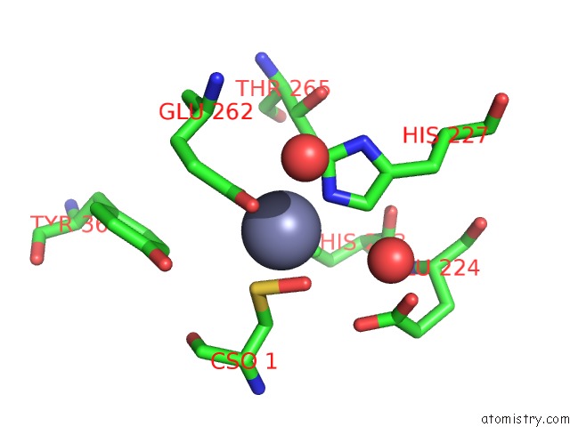
Mono view
Stereo pair view
Mono view
Stereo pair view
A full contact list of Zinc with other atoms in the Zn binding
site number 1 of Structure of the C. Botulinum Neurotoxin Serotype A with An Inhibitory Peptide Bound within 5.0Å range:
|
Reference:
N.R.Silvaggi,
D.Wilson,
S.Tzipori,
K.N.Allen.
Catalytic Features of the Botulinum Neurotoxin A Light Chain Revealed By High Resolution Structure of An Inhibitory Peptide Complex. Biochemistry V. 47 5736 2008.
ISSN: ISSN 0006-2960
PubMed: 18457419
DOI: 10.1021/BI8001067
Page generated: Wed Aug 20 08:03:33 2025
ISSN: ISSN 0006-2960
PubMed: 18457419
DOI: 10.1021/BI8001067
Last articles
Zn in 3RZUZn in 3S0Z
Zn in 3RZO
Zn in 3RZD
Zn in 3S0N
Zn in 3RZV
Zn in 3RYM
Zn in 3RZA
Zn in 3RZ8
Zn in 3RZ7