Zinc »
PDB 3b7r-3bo5 »
3b7t »
Zinc in PDB 3b7t: [E296Q]LTA4H in Complex with Arg-Ala-Arg Substrate
Enzymatic activity of [E296Q]LTA4H in Complex with Arg-Ala-Arg Substrate
All present enzymatic activity of [E296Q]LTA4H in Complex with Arg-Ala-Arg Substrate:
3.3.2.6;
3.3.2.6;
Protein crystallography data
The structure of [E296Q]LTA4H in Complex with Arg-Ala-Arg Substrate, PDB code: 3b7t
was solved by
F.Tholander,
J.Haeggstrom,
M.Thunnissen,
A.Muroya,
B.-P.Roques,
M.-C.Fournie-Zaluski,
with X-Ray Crystallography technique. A brief refinement statistics is given in the table below:
| Resolution Low / High (Å) | 30.00 / 2.30 |
| Space group | P 21 21 21 |
| Cell size a, b, c (Å), α, β, γ (°) | 78.490, 87.785, 99.916, 90.00, 90.00, 90.00 |
| R / Rfree (%) | 20.6 / 27.3 |
Other elements in 3b7t:
The structure of [E296Q]LTA4H in Complex with Arg-Ala-Arg Substrate also contains other interesting chemical elements:
| Ytterbium | (Yb) | 2 atoms |
Zinc Binding Sites:
The binding sites of Zinc atom in the [E296Q]LTA4H in Complex with Arg-Ala-Arg Substrate
(pdb code 3b7t). This binding sites where shown within
5.0 Angstroms radius around Zinc atom.
In total only one binding site of Zinc was determined in the [E296Q]LTA4H in Complex with Arg-Ala-Arg Substrate, PDB code: 3b7t:
In total only one binding site of Zinc was determined in the [E296Q]LTA4H in Complex with Arg-Ala-Arg Substrate, PDB code: 3b7t:
Zinc binding site 1 out of 1 in 3b7t
Go back to
Zinc binding site 1 out
of 1 in the [E296Q]LTA4H in Complex with Arg-Ala-Arg Substrate
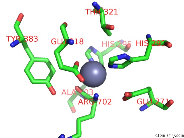
Mono view
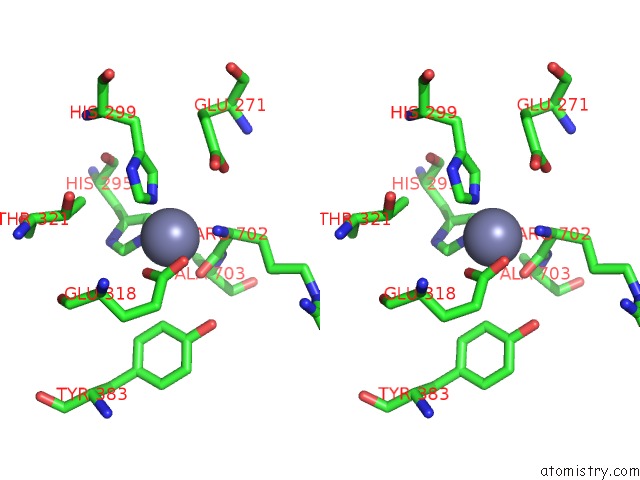
Stereo pair view
Mono view
Stereo pair view
A full contact list of Zinc with other atoms in the Zn binding
site number 1 of [E296Q]LTA4H in Complex with Arg-Ala-Arg Substrate within 5.0Å range:
|
Reference:
F.Tholander,
A.Muroya,
B.P.Roques,
M.C.Fournie-Zaluski,
M.M.Thunnissen,
J.Z.Haeggstrom.
Structure-Based Dissection of the Active Site Chemistry of Leukotriene A4 Hydrolase: Implications For M1 Aminopeptidases and Inhibitor Design. Chem.Biol. V. 15 920 2008.
ISSN: ISSN 1074-5521
PubMed: 18804029
DOI: 10.1016/J.CHEMBIOL.2008.07.018
Page generated: Thu Oct 24 11:27:30 2024
ISSN: ISSN 1074-5521
PubMed: 18804029
DOI: 10.1016/J.CHEMBIOL.2008.07.018
Last articles
Zn in 9MJ5Zn in 9HNW
Zn in 9G0L
Zn in 9FNE
Zn in 9DZN
Zn in 9E0I
Zn in 9D32
Zn in 9DAK
Zn in 8ZXC
Zn in 8ZUF