Zinc »
PDB 2zep-2zxg »
2znr »
Zinc in PDB 2znr: Crystal Structure of the Dub Domain of Human Amsh-Lp
Enzymatic activity of Crystal Structure of the Dub Domain of Human Amsh-Lp
All present enzymatic activity of Crystal Structure of the Dub Domain of Human Amsh-Lp:
3.1.2.15;
3.1.2.15;
Protein crystallography data
The structure of Crystal Structure of the Dub Domain of Human Amsh-Lp, PDB code: 2znr
was solved by
Y.Sato,
Y.Azusa,
A.Yamagata,
H.Mimura,
X.Wang,
M.Yamashita,
K.Ookata,
O.Nureki,
K.Iwai,
M.Komada,
S.Fukai,
with X-Ray Crystallography technique. A brief refinement statistics is given in the table below:
| Resolution Low / High (Å) | 31.09 / 1.20 |
| Space group | P 65 |
| Cell size a, b, c (Å), α, β, γ (°) | 81.865, 81.865, 64.693, 90.00, 90.00, 120.00 |
| R / Rfree (%) | 14.9 / 16.5 |
Other elements in 2znr:
The structure of Crystal Structure of the Dub Domain of Human Amsh-Lp also contains other interesting chemical elements:
| Praseodymium | (Pr) | 1 atom |
Zinc Binding Sites:
The binding sites of Zinc atom in the Crystal Structure of the Dub Domain of Human Amsh-Lp
(pdb code 2znr). This binding sites where shown within
5.0 Angstroms radius around Zinc atom.
In total 2 binding sites of Zinc where determined in the Crystal Structure of the Dub Domain of Human Amsh-Lp, PDB code: 2znr:
Jump to Zinc binding site number: 1; 2;
In total 2 binding sites of Zinc where determined in the Crystal Structure of the Dub Domain of Human Amsh-Lp, PDB code: 2znr:
Jump to Zinc binding site number: 1; 2;
Zinc binding site 1 out of 2 in 2znr
Go back to
Zinc binding site 1 out
of 2 in the Crystal Structure of the Dub Domain of Human Amsh-Lp
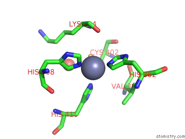
Mono view
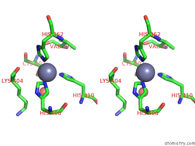
Stereo pair view
Mono view
Stereo pair view
A full contact list of Zinc with other atoms in the Zn binding
site number 1 of Crystal Structure of the Dub Domain of Human Amsh-Lp within 5.0Å range:
|
Zinc binding site 2 out of 2 in 2znr
Go back to
Zinc binding site 2 out
of 2 in the Crystal Structure of the Dub Domain of Human Amsh-Lp
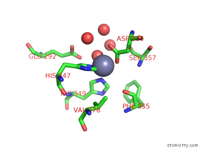
Mono view
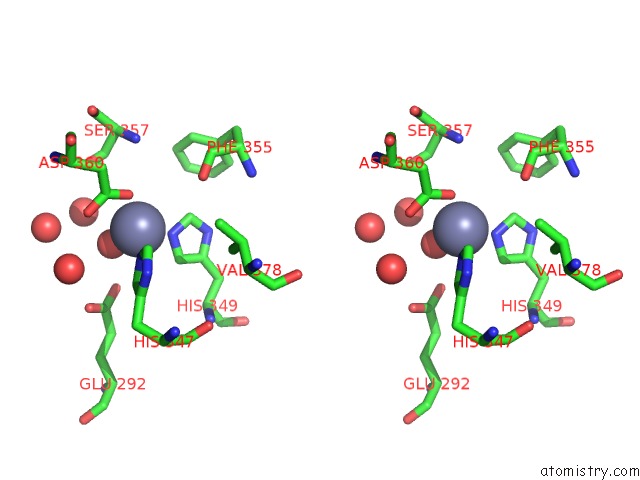
Stereo pair view
Mono view
Stereo pair view
A full contact list of Zinc with other atoms in the Zn binding
site number 2 of Crystal Structure of the Dub Domain of Human Amsh-Lp within 5.0Å range:
|
Reference:
Y.Sato,
A.Yoshikawa,
A.Yamagata,
H.Mimura,
M.Yamashita,
K.Ookata,
O.Nureki,
K.Iwai,
M.Komada,
S.Fukai.
Structural Basis For Specific Cleavage of Lys 63-Linked Polyubiquitin Chains Nature V. 455 358 2008.
ISSN: ISSN 0028-0836
PubMed: 18758443
DOI: 10.1038/NATURE07254
Page generated: Thu Oct 24 10:50:36 2024
ISSN: ISSN 0028-0836
PubMed: 18758443
DOI: 10.1038/NATURE07254
Last articles
Zn in 9MJ5Zn in 9HNW
Zn in 9G0L
Zn in 9FNE
Zn in 9DZN
Zn in 9E0I
Zn in 9D32
Zn in 9DAK
Zn in 8ZXC
Zn in 8ZUF