Zinc »
PDB 2y7g-2yqp »
2yd0 »
Zinc in PDB 2yd0: Crystal Structure of the Soluble Domain of Human Endoplasmic Reticulum Aminopeptidase 1 ERAP1
Protein crystallography data
The structure of Crystal Structure of the Soluble Domain of Human Endoplasmic Reticulum Aminopeptidase 1 ERAP1, PDB code: 2yd0
was solved by
M.Vollmar,
G.Kochan,
T.Krojer,
E.Ugochukwu,
J.R.C.Muniz,
J.Raynor,
A.Chaikuad,
C.Allerston,
F.Von Delft,
C.Bountra,
C.H.Arrowsmith,
J.Weigelt,
A.Edwards,
S.Knapp,
with X-Ray Crystallography technique. A brief refinement statistics is given in the table below:
| Resolution Low / High (Å) | 19.94 / 2.70 |
| Space group | P 6 2 2 |
| Cell size a, b, c (Å), α, β, γ (°) | 200.948, 200.948, 114.222, 90.00, 90.00, 120.00 |
| R / Rfree (%) | 15.2 / 21.5 |
Other elements in 2yd0:
The structure of Crystal Structure of the Soluble Domain of Human Endoplasmic Reticulum Aminopeptidase 1 ERAP1 also contains other interesting chemical elements:
| Potassium | (K) | 2 atoms |
Zinc Binding Sites:
The binding sites of Zinc atom in the Crystal Structure of the Soluble Domain of Human Endoplasmic Reticulum Aminopeptidase 1 ERAP1
(pdb code 2yd0). This binding sites where shown within
5.0 Angstroms radius around Zinc atom.
In total only one binding site of Zinc was determined in the Crystal Structure of the Soluble Domain of Human Endoplasmic Reticulum Aminopeptidase 1 ERAP1, PDB code: 2yd0:
In total only one binding site of Zinc was determined in the Crystal Structure of the Soluble Domain of Human Endoplasmic Reticulum Aminopeptidase 1 ERAP1, PDB code: 2yd0:
Zinc binding site 1 out of 1 in 2yd0
Go back to
Zinc binding site 1 out
of 1 in the Crystal Structure of the Soluble Domain of Human Endoplasmic Reticulum Aminopeptidase 1 ERAP1
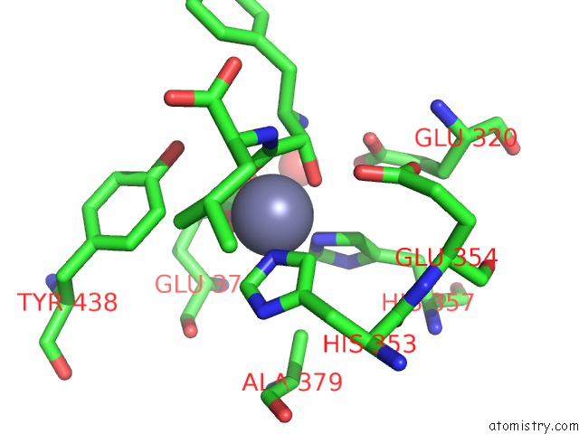
Mono view
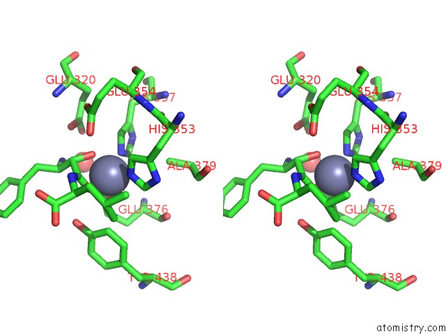
Stereo pair view
Mono view
Stereo pair view
A full contact list of Zinc with other atoms in the Zn binding
site number 1 of Crystal Structure of the Soluble Domain of Human Endoplasmic Reticulum Aminopeptidase 1 ERAP1 within 5.0Å range:
|
Reference:
G.Kochan,
T.Krojer,
D.Harvey,
R.Fischer,
L.Chen,
M.Vollmar,
F.Von Delft,
K.L.Kavanagh,
M.A.Brown,
P.Bowness,
P.Wordsworth,
B.M.Kessler,
U.Oppermann.
Crystal Structures of the Endoplasmic Reticulum Aminopeptidase-1 (ERAP1) Reveal the Molecular Basis For N-Terminal Peptide Trimming. Proc.Natl.Acad.Sci.Usa V. 108 7745 2011.
ISSN: ISSN 0027-8424
PubMed: 21508329
DOI: 10.1073/PNAS.1101262108
Page generated: Thu Oct 17 05:49:20 2024
ISSN: ISSN 0027-8424
PubMed: 21508329
DOI: 10.1073/PNAS.1101262108
Last articles
Zn in 9MJ5Zn in 9HNW
Zn in 9G0L
Zn in 9FNE
Zn in 9DZN
Zn in 9E0I
Zn in 9D32
Zn in 9DAK
Zn in 8ZXC
Zn in 8ZUF