Zinc »
PDB 2x91-2xjz »
2xgw »
Zinc in PDB 2xgw: Zinc-Bound Crystal Structure of Streptococcus Pyogenes Dpr
Protein crystallography data
The structure of Zinc-Bound Crystal Structure of Streptococcus Pyogenes Dpr, PDB code: 2xgw
was solved by
T.Haikarainen,
C.-C.Tsou,
J.-J.Wu,
A.C.Papageorgiou,
with X-Ray Crystallography technique. A brief refinement statistics is given in the table below:
| Resolution Low / High (Å) | 107.83 / 2.10 |
| Space group | F 41 3 2 |
| Cell size a, b, c (Å), α, β, γ (°) | 187.120, 187.120, 187.120, 90.00, 90.00, 90.00 |
| R / Rfree (%) | 14.4 / 17.2 |
Other elements in 2xgw:
The structure of Zinc-Bound Crystal Structure of Streptococcus Pyogenes Dpr also contains other interesting chemical elements:
| Chlorine | (Cl) | 1 atom |
Zinc Binding Sites:
The binding sites of Zinc atom in the Zinc-Bound Crystal Structure of Streptococcus Pyogenes Dpr
(pdb code 2xgw). This binding sites where shown within
5.0 Angstroms radius around Zinc atom.
In total 4 binding sites of Zinc where determined in the Zinc-Bound Crystal Structure of Streptococcus Pyogenes Dpr, PDB code: 2xgw:
Jump to Zinc binding site number: 1; 2; 3; 4;
In total 4 binding sites of Zinc where determined in the Zinc-Bound Crystal Structure of Streptococcus Pyogenes Dpr, PDB code: 2xgw:
Jump to Zinc binding site number: 1; 2; 3; 4;
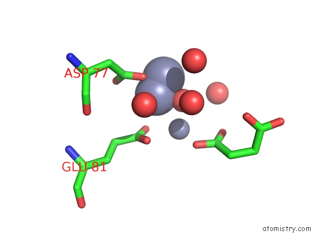
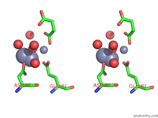
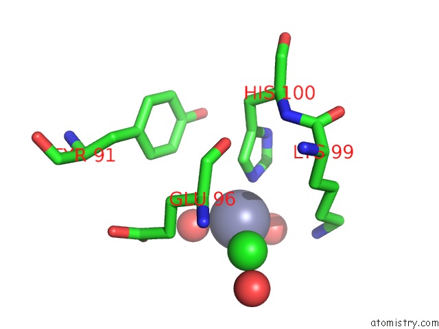
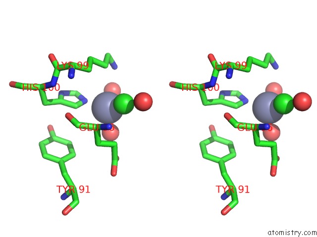
Zinc binding site 1 out of 4 in 2xgw
Go back to
Zinc binding site 1 out
of 4 in the Zinc-Bound Crystal Structure of Streptococcus Pyogenes Dpr

Mono view

Stereo pair view
Mono view
Stereo pair view
A full contact list of Zinc with other atoms in the Zn binding
site number 1 of Zinc-Bound Crystal Structure of Streptococcus Pyogenes Dpr within 5.0Å range:
|
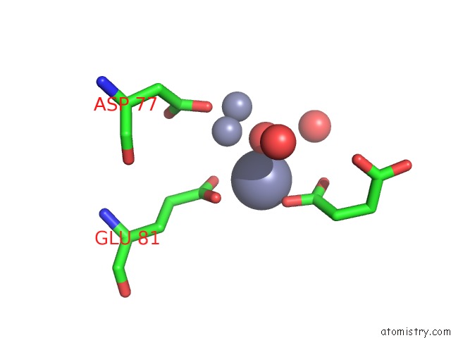
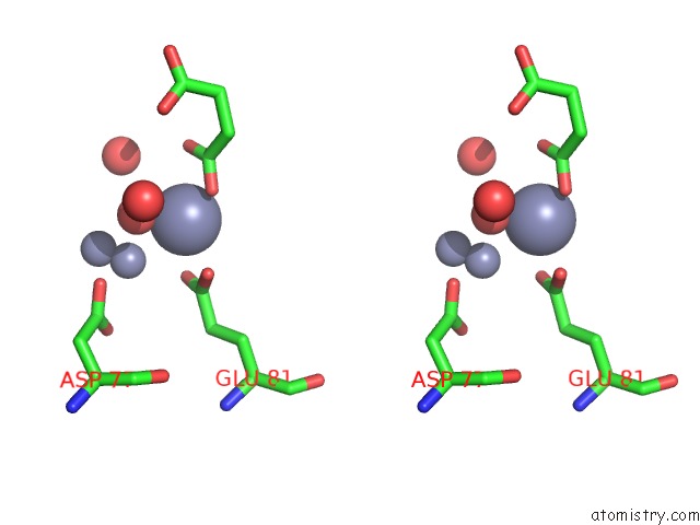
Zinc binding site 2 out of 4 in 2xgw
Go back to
Zinc binding site 2 out
of 4 in the Zinc-Bound Crystal Structure of Streptococcus Pyogenes Dpr
Mono view
Stereo pair view
Mono view
Stereo pair view
A full contact list of Zinc with other atoms in the Zn binding
site number 2 of Zinc-Bound Crystal Structure of Streptococcus Pyogenes Dpr within 5.0Å range:
|
Zinc binding site 3 out of 4 in 2xgw
Go back to
Zinc binding site 3 out
of 4 in the Zinc-Bound Crystal Structure of Streptococcus Pyogenes Dpr
Mono view
Stereo pair view
Mono view
Stereo pair view
A full contact list of Zinc with other atoms in the Zn binding
site number 3 of Zinc-Bound Crystal Structure of Streptococcus Pyogenes Dpr within 5.0Å range:
|
Zinc binding site 4 out of 4 in 2xgw
Go back to
Zinc binding site 4 out
of 4 in the Zinc-Bound Crystal Structure of Streptococcus Pyogenes Dpr
Mono view
Stereo pair view
Mono view
Stereo pair view
A full contact list of Zinc with other atoms in the Zn binding
site number 4 of Zinc-Bound Crystal Structure of Streptococcus Pyogenes Dpr within 5.0Å range:
|
Reference:
T.Haikarainen,
C.-C.Tsou,
J.-J.Wu,
A.C.Papageorgiou.
Structural Characterization and Biological Implications of Di-Zinc Binding in the Ferroxidase Center of Streptococcus Pyogenes Dpr. Biochem.Biophys.Res.Commun. V. 398 361 2010.
ISSN: ISSN 0006-291X
PubMed: 20599728
DOI: 10.1016/J.BBRC.2010.06.071
Page generated: Thu Oct 17 05:18:22 2024
ISSN: ISSN 0006-291X
PubMed: 20599728
DOI: 10.1016/J.BBRC.2010.06.071
Last articles
Zn in 9MJ5Zn in 9HNW
Zn in 9G0L
Zn in 9FNE
Zn in 9DZN
Zn in 9E0I
Zn in 9D32
Zn in 9DAK
Zn in 8ZXC
Zn in 8ZUF