Zinc »
PDB 2wx0-2x91 »
2x0w »
Zinc in PDB 2x0w: Structure of the P53 Core Domain Mutant Y220C Bound to 5,6-Dimethoxy- 2-Methylbenzothiazole
Protein crystallography data
The structure of Structure of the P53 Core Domain Mutant Y220C Bound to 5,6-Dimethoxy- 2-Methylbenzothiazole, PDB code: 2x0w
was solved by
J.L.Kaar,
N.Basse,
A.C.Joerger,
A.R.Fersht,
with X-Ray Crystallography technique. A brief refinement statistics is given in the table below:
| Resolution Low / High (Å) | 24.90 / 2.10 |
| Space group | P 21 21 21 |
| Cell size a, b, c (Å), α, β, γ (°) | 65.250, 70.580, 105.440, 90.00, 90.00, 90.00 |
| R / Rfree (%) | 17.2 / 22.8 |
Zinc Binding Sites:
The binding sites of Zinc atom in the Structure of the P53 Core Domain Mutant Y220C Bound to 5,6-Dimethoxy- 2-Methylbenzothiazole
(pdb code 2x0w). This binding sites where shown within
5.0 Angstroms radius around Zinc atom.
In total 2 binding sites of Zinc where determined in the Structure of the P53 Core Domain Mutant Y220C Bound to 5,6-Dimethoxy- 2-Methylbenzothiazole, PDB code: 2x0w:
Jump to Zinc binding site number: 1; 2;
In total 2 binding sites of Zinc where determined in the Structure of the P53 Core Domain Mutant Y220C Bound to 5,6-Dimethoxy- 2-Methylbenzothiazole, PDB code: 2x0w:
Jump to Zinc binding site number: 1; 2;
Zinc binding site 1 out of 2 in 2x0w
Go back to
Zinc binding site 1 out
of 2 in the Structure of the P53 Core Domain Mutant Y220C Bound to 5,6-Dimethoxy- 2-Methylbenzothiazole
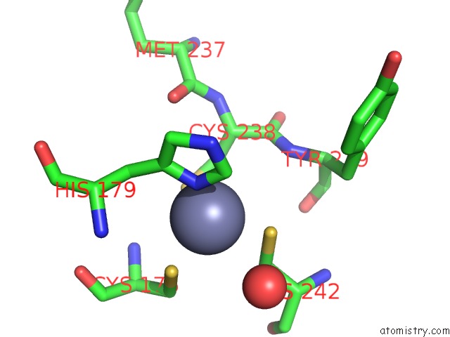
Mono view
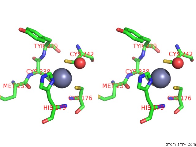
Stereo pair view
Mono view
Stereo pair view
A full contact list of Zinc with other atoms in the Zn binding
site number 1 of Structure of the P53 Core Domain Mutant Y220C Bound to 5,6-Dimethoxy- 2-Methylbenzothiazole within 5.0Å range:
|
Zinc binding site 2 out of 2 in 2x0w
Go back to
Zinc binding site 2 out
of 2 in the Structure of the P53 Core Domain Mutant Y220C Bound to 5,6-Dimethoxy- 2-Methylbenzothiazole
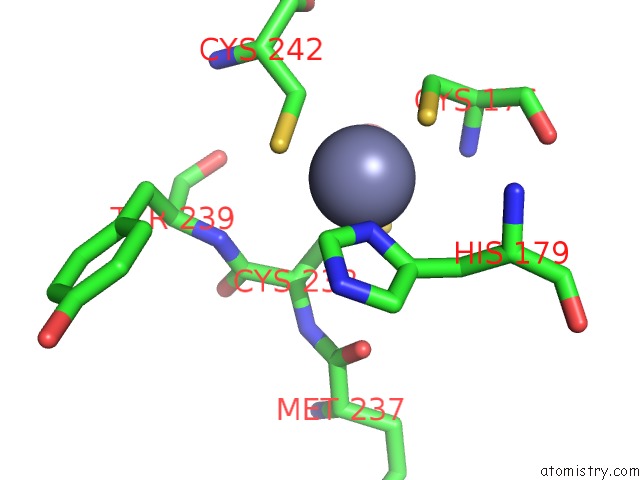
Mono view
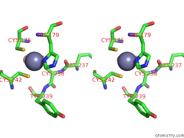
Stereo pair view
Mono view
Stereo pair view
A full contact list of Zinc with other atoms in the Zn binding
site number 2 of Structure of the P53 Core Domain Mutant Y220C Bound to 5,6-Dimethoxy- 2-Methylbenzothiazole within 5.0Å range:
|
Reference:
N.Basse,
J.L.Kaar,
G.Settanni,
A.C.Joerger,
T.J.Rutherford,
A.R.Fersht.
Toward the Rational Design of P53-Stabilizing Drugs: Probing the Surface of the Oncogenic Y220C Mutant. Chem.Biol. V. 17 46 2010.
ISSN: ISSN 1074-5521
PubMed: 20142040
DOI: 10.1016/J.CHEMBIOL.2009.12.011
Page generated: Wed Aug 20 06:34:52 2025
ISSN: ISSN 1074-5521
PubMed: 20142040
DOI: 10.1016/J.CHEMBIOL.2009.12.011
Last articles
Zn in 3QN9Zn in 3QMG
Zn in 3QMH
Zn in 3QMI
Zn in 3QMD
Zn in 3QM3
Zn in 3QLN
Zn in 3QMC
Zn in 3QMB
Zn in 3QLC