Zinc »
PDB 2wfx-2wwy »
2wor »
Zinc in PDB 2wor: Co-Structure of S100A7 with 1,8 Ans
Protein crystallography data
The structure of Co-Structure of S100A7 with 1,8 Ans, PDB code: 2wor
was solved by
R.Leon,
F.Hof,
M.J.Boulanger,
with X-Ray Crystallography technique. A brief refinement statistics is given in the table below:
| Resolution Low / High (Å) | 47.27 / 1.70 |
| Space group | P 43 21 2 |
| Cell size a, b, c (Å), α, β, γ (°) | 51.660, 51.660, 117.240, 90.00, 90.00, 90.00 |
| R / Rfree (%) | 18.673 / 21.211 |
Other elements in 2wor:
The structure of Co-Structure of S100A7 with 1,8 Ans also contains other interesting chemical elements:
| Calcium | (Ca) | 1 atom |
Zinc Binding Sites:
The binding sites of Zinc atom in the Co-Structure of S100A7 with 1,8 Ans
(pdb code 2wor). This binding sites where shown within
5.0 Angstroms radius around Zinc atom.
In total only one binding site of Zinc was determined in the Co-Structure of S100A7 with 1,8 Ans, PDB code: 2wor:
In total only one binding site of Zinc was determined in the Co-Structure of S100A7 with 1,8 Ans, PDB code: 2wor:
Zinc binding site 1 out of 1 in 2wor
Go back to
Zinc binding site 1 out
of 1 in the Co-Structure of S100A7 with 1,8 Ans
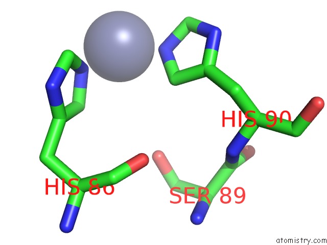
Mono view
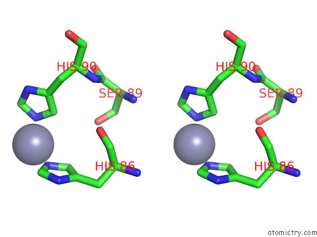
Stereo pair view
Mono view
Stereo pair view
A full contact list of Zinc with other atoms in the Zn binding
site number 1 of Co-Structure of S100A7 with 1,8 Ans within 5.0Å range:
|
Reference:
R.Leon,
J.I.Murray,
G.Cragg,
B.Farnell,
N.R.West,
T.C.Pace,
P.H.Watson,
C.Bohne,
M.J.Boulanger,
F.Hof.
Identification and Characterization of Binding Sites on S100A7, A Participant in Cancer and Inflammation Pathways. Biochemistry V. 48 10591 2009.
ISSN: ISSN 0006-2960
PubMed: 19810752
DOI: 10.1021/BI901330G
Page generated: Thu Oct 17 04:59:56 2024
ISSN: ISSN 0006-2960
PubMed: 19810752
DOI: 10.1021/BI901330G
Last articles
Zn in 9MJ5Zn in 9HNW
Zn in 9G0L
Zn in 9FNE
Zn in 9DZN
Zn in 9E0I
Zn in 9D32
Zn in 9DAK
Zn in 8ZXC
Zn in 8ZUF