Zinc »
PDB 2wfx-2wwy »
2wkx »
Zinc in PDB 2wkx: Crystal Structure of the Native E. Coli Zinc Amidase Amid
Enzymatic activity of Crystal Structure of the Native E. Coli Zinc Amidase Amid
All present enzymatic activity of Crystal Structure of the Native E. Coli Zinc Amidase Amid:
3.5.1.28;
3.5.1.28;
Protein crystallography data
The structure of Crystal Structure of the Native E. Coli Zinc Amidase Amid, PDB code: 2wkx
was solved by
S.Petrella,
F.Kerff,
R.Herman,
C.Genereux,
A.Pennartz,
E.Sauvage,
B.Joris,
P.Charlier,
with X-Ray Crystallography technique. A brief refinement statistics is given in the table below:
| Resolution Low / High (Å) | 21.23 / 1.80 |
| Space group | P 61 2 2 |
| Cell size a, b, c (Å), α, β, γ (°) | 88.990, 88.990, 183.923, 90.00, 90.00, 120.00 |
| R / Rfree (%) | 16.077 / 18.367 |
Other elements in 2wkx:
The structure of Crystal Structure of the Native E. Coli Zinc Amidase Amid also contains other interesting chemical elements:
| Chlorine | (Cl) | 2 atoms |
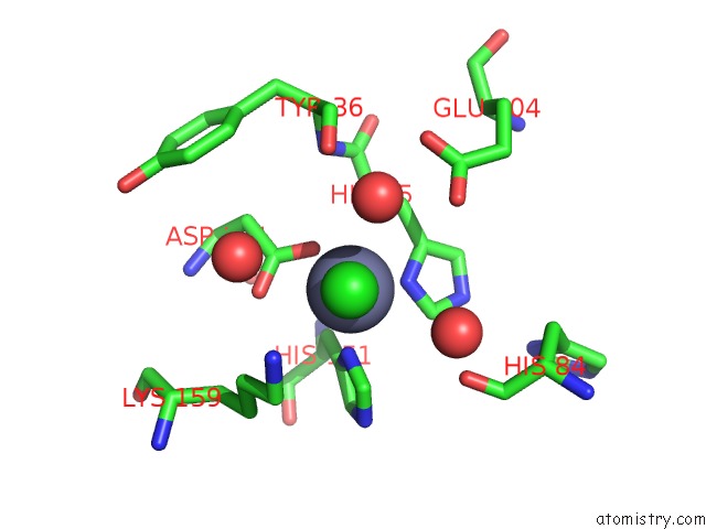
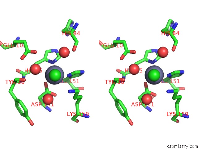
Zinc Binding Sites:
The binding sites of Zinc atom in the Crystal Structure of the Native E. Coli Zinc Amidase Amid
(pdb code 2wkx). This binding sites where shown within
5.0 Angstroms radius around Zinc atom.
In total only one binding site of Zinc was determined in the Crystal Structure of the Native E. Coli Zinc Amidase Amid, PDB code: 2wkx:
In total only one binding site of Zinc was determined in the Crystal Structure of the Native E. Coli Zinc Amidase Amid, PDB code: 2wkx:
Zinc binding site 1 out of 1 in 2wkx
Go back to
Zinc binding site 1 out
of 1 in the Crystal Structure of the Native E. Coli Zinc Amidase Amid
Mono view
Stereo pair view
Mono view
Stereo pair view
A full contact list of Zinc with other atoms in the Zn binding
site number 1 of Crystal Structure of the Native E. Coli Zinc Amidase Amid within 5.0Å range:
|
Reference:
F.Kerff,
S.Petrella,
F.Mercier,
E.Sauvage,
R.Herman,
A.Pennartz,
A.Zervosen,
A.Luxen,
J.M.Frere,
B.Joris,
P.Charlier.
Specific Structural Features of the N- Acetylmuramoyl-L-Alanine Amidase Amid From Escherichia Coli and Mechanistic Implications For Enzymes of This Family. J.Mol.Biol. V. 397 249 2010.
ISSN: ISSN 0022-2836
PubMed: 20036252
DOI: 10.1016/J.JMB.2009.12.038
Page generated: Thu Oct 17 04:56:31 2024
ISSN: ISSN 0022-2836
PubMed: 20036252
DOI: 10.1016/J.JMB.2009.12.038
Last articles
Zn in 9MJ5Zn in 9HNW
Zn in 9G0L
Zn in 9FNE
Zn in 9DZN
Zn in 9E0I
Zn in 9D32
Zn in 9DAK
Zn in 8ZXC
Zn in 8ZUF