Zinc »
PDB 2vhf-2vr2 »
2vmk »
Zinc in PDB 2vmk: Crystal Structure of E. Coli Rnase E Apoprotein - Catalytic Domain
Protein crystallography data
The structure of Crystal Structure of E. Coli Rnase E Apoprotein - Catalytic Domain, PDB code: 2vmk
was solved by
D.J.Koslover,
A.J.Callaghan,
M.J.Marcaida,
M.Martick,
W.G.Scott,
B.F.Luisi,
with X-Ray Crystallography technique. A brief refinement statistics is given in the table below:
| Resolution Low / High (Å) | 47.84 / 3.3 |
| Space group | P 1 |
| Cell size a, b, c (Å), α, β, γ (°) | 73.241, 75.571, 109.370, 94.95, 102.03, 91.77 |
| R / Rfree (%) | 26.7 / 29.3 |
Zinc Binding Sites:
The binding sites of Zinc atom in the Crystal Structure of E. Coli Rnase E Apoprotein - Catalytic Domain
(pdb code 2vmk). This binding sites where shown within
5.0 Angstroms radius around Zinc atom.
In total 2 binding sites of Zinc where determined in the Crystal Structure of E. Coli Rnase E Apoprotein - Catalytic Domain, PDB code: 2vmk:
Jump to Zinc binding site number: 1; 2;
In total 2 binding sites of Zinc where determined in the Crystal Structure of E. Coli Rnase E Apoprotein - Catalytic Domain, PDB code: 2vmk:
Jump to Zinc binding site number: 1; 2;
Zinc binding site 1 out of 2 in 2vmk
Go back to
Zinc binding site 1 out
of 2 in the Crystal Structure of E. Coli Rnase E Apoprotein - Catalytic Domain
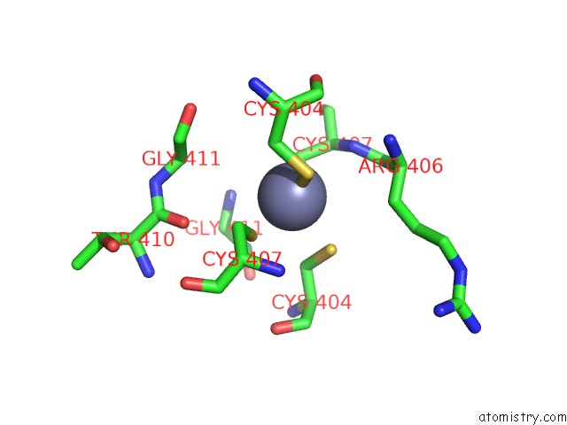
Mono view
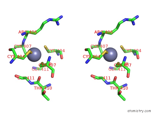
Stereo pair view
Mono view
Stereo pair view
A full contact list of Zinc with other atoms in the Zn binding
site number 1 of Crystal Structure of E. Coli Rnase E Apoprotein - Catalytic Domain within 5.0Å range:
|
Zinc binding site 2 out of 2 in 2vmk
Go back to
Zinc binding site 2 out
of 2 in the Crystal Structure of E. Coli Rnase E Apoprotein - Catalytic Domain
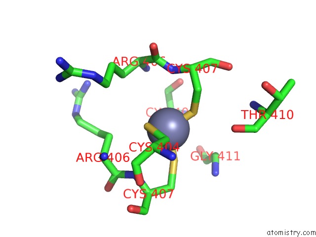
Mono view
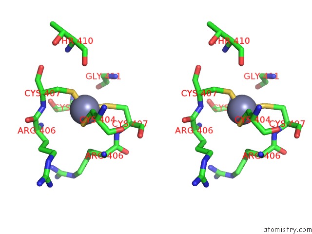
Stereo pair view
Mono view
Stereo pair view
A full contact list of Zinc with other atoms in the Zn binding
site number 2 of Crystal Structure of E. Coli Rnase E Apoprotein - Catalytic Domain within 5.0Å range:
|
Reference:
D.J.Koslover,
A.J.Callaghan,
M.J.Marcaida,
E.F.Garman,
M.Martick,
W.G.Scott,
B.F.Luisi.
The Crystal Structure of the Escherichia Coli Rnase E Apoprotein and A Mechanism For Rna Degradation. Structure V. 16 1238 2008.
ISSN: ISSN 0969-2126
PubMed: 18682225
DOI: 10.1016/J.STR.2008.04.017
Page generated: Thu Oct 17 04:23:07 2024
ISSN: ISSN 0969-2126
PubMed: 18682225
DOI: 10.1016/J.STR.2008.04.017
Last articles
Zn in 9MJ5Zn in 9HNW
Zn in 9G0L
Zn in 9FNE
Zn in 9DZN
Zn in 9E0I
Zn in 9D32
Zn in 9DAK
Zn in 8ZXC
Zn in 8ZUF