Zinc »
PDB 2vhf-2vr2 »
2vhf »
Zinc in PDB 2vhf: Structure of the Cyld Usp Domain
Enzymatic activity of Structure of the Cyld Usp Domain
All present enzymatic activity of Structure of the Cyld Usp Domain:
3.1.2.15;
3.1.2.15;
Protein crystallography data
The structure of Structure of the Cyld Usp Domain, PDB code: 2vhf
was solved by
D.Komander,
C.J.Lord,
H.Scheel,
S.Swift,
K.Hofmann,
A.Ashworth,
D.Barford,
with X-Ray Crystallography technique. A brief refinement statistics is given in the table below:
| Resolution Low / High (Å) | 29.79 / 2.8 |
| Space group | P 21 21 21 |
| Cell size a, b, c (Å), α, β, γ (°) | 60.494, 89.083, 171.806, 90.00, 90.00, 90.00 |
| R / Rfree (%) | 23.3 / 28.1 |
Zinc Binding Sites:
The binding sites of Zinc atom in the Structure of the Cyld Usp Domain
(pdb code 2vhf). This binding sites where shown within
5.0 Angstroms radius around Zinc atom.
In total 4 binding sites of Zinc where determined in the Structure of the Cyld Usp Domain, PDB code: 2vhf:
Jump to Zinc binding site number: 1; 2; 3; 4;
In total 4 binding sites of Zinc where determined in the Structure of the Cyld Usp Domain, PDB code: 2vhf:
Jump to Zinc binding site number: 1; 2; 3; 4;
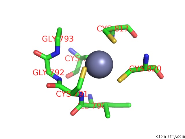
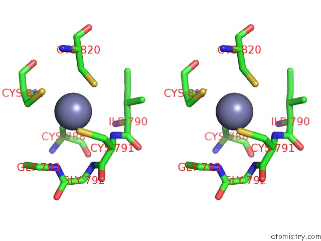
Zinc binding site 1 out of 4 in 2vhf
Go back to
Zinc binding site 1 out
of 4 in the Structure of the Cyld Usp Domain
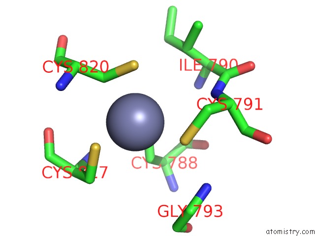
Mono view
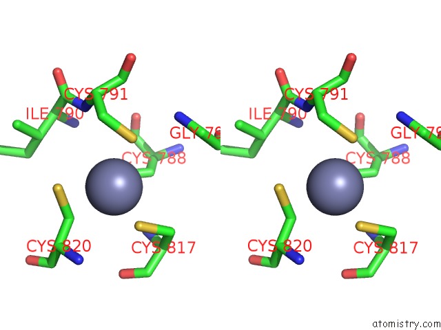
Stereo pair view
Mono view
Stereo pair view
A full contact list of Zinc with other atoms in the Zn binding
site number 1 of Structure of the Cyld Usp Domain within 5.0Å range:
|
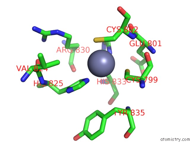
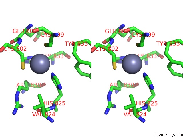
Zinc binding site 2 out of 4 in 2vhf
Go back to
Zinc binding site 2 out
of 4 in the Structure of the Cyld Usp Domain
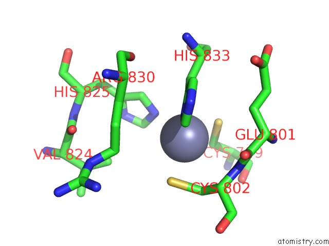
Mono view
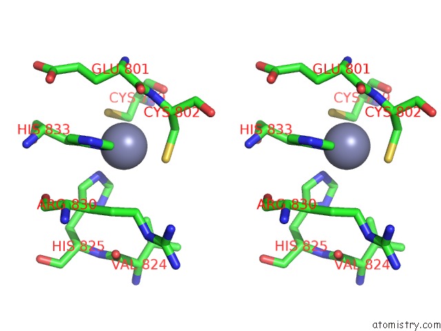
Stereo pair view
Mono view
Stereo pair view
A full contact list of Zinc with other atoms in the Zn binding
site number 2 of Structure of the Cyld Usp Domain within 5.0Å range:
|
Zinc binding site 3 out of 4 in 2vhf
Go back to
Zinc binding site 3 out
of 4 in the Structure of the Cyld Usp Domain
Mono view
Stereo pair view
Mono view
Stereo pair view
A full contact list of Zinc with other atoms in the Zn binding
site number 3 of Structure of the Cyld Usp Domain within 5.0Å range:
|
Zinc binding site 4 out of 4 in 2vhf
Go back to
Zinc binding site 4 out
of 4 in the Structure of the Cyld Usp Domain
Mono view
Stereo pair view
Mono view
Stereo pair view
A full contact list of Zinc with other atoms in the Zn binding
site number 4 of Structure of the Cyld Usp Domain within 5.0Å range:
|
Reference:
D.Komander,
C.J.Lord,
H.Scheel,
S.Swift,
K.Hofmann,
A.Ashworth,
D.Barford.
The Structure of the Cyld Usp Domain Explains Its Specificity For LYS63-Linked Polyubiquitin and Reveals A B-Box Module Mol.Cell.Biol. V. 29 451 2008.
ISSN: ISSN 0270-7306
PubMed: 18313383
DOI: 10.1016/J.MOLCEL.2007.12.018
Page generated: Thu Oct 17 04:18:32 2024
ISSN: ISSN 0270-7306
PubMed: 18313383
DOI: 10.1016/J.MOLCEL.2007.12.018
Last articles
Zn in 9MJ5Zn in 9HNW
Zn in 9G0L
Zn in 9FNE
Zn in 9DZN
Zn in 9E0I
Zn in 9D32
Zn in 9DAK
Zn in 8ZXC
Zn in 8ZUF