Zinc »
PDB 2v20-2vh9 »
2ves »
Zinc in PDB 2ves: Crystal Structure of Lpxc From Pseudomonas Aeruginosa Complexed with the Potent Bb-78485 Inhibitor
Enzymatic activity of Crystal Structure of Lpxc From Pseudomonas Aeruginosa Complexed with the Potent Bb-78485 Inhibitor
All present enzymatic activity of Crystal Structure of Lpxc From Pseudomonas Aeruginosa Complexed with the Potent Bb-78485 Inhibitor:
3.5.1.108;
3.5.1.108;
Protein crystallography data
The structure of Crystal Structure of Lpxc From Pseudomonas Aeruginosa Complexed with the Potent Bb-78485 Inhibitor, PDB code: 2ves
was solved by
I.Mochalkin,
J.D.Knafels,
with X-Ray Crystallography technique. A brief refinement statistics is given in the table below:
| Resolution Low / High (Å) | 73.72 / 1.90 |
| Space group | P 21 21 21 |
| Cell size a, b, c (Å), α, β, γ (°) | 90.542, 103.331, 105.284, 90.00, 90.00, 90.00 |
| R / Rfree (%) | 17.7 / 22.1 |
Zinc Binding Sites:
The binding sites of Zinc atom in the Crystal Structure of Lpxc From Pseudomonas Aeruginosa Complexed with the Potent Bb-78485 Inhibitor
(pdb code 2ves). This binding sites where shown within
5.0 Angstroms radius around Zinc atom.
In total 7 binding sites of Zinc where determined in the Crystal Structure of Lpxc From Pseudomonas Aeruginosa Complexed with the Potent Bb-78485 Inhibitor, PDB code: 2ves:
Jump to Zinc binding site number: 1; 2; 3; 4; 5; 6; 7;
In total 7 binding sites of Zinc where determined in the Crystal Structure of Lpxc From Pseudomonas Aeruginosa Complexed with the Potent Bb-78485 Inhibitor, PDB code: 2ves:
Jump to Zinc binding site number: 1; 2; 3; 4; 5; 6; 7;
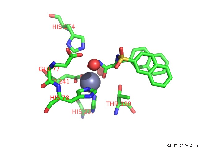
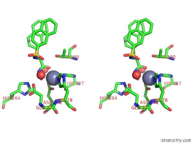
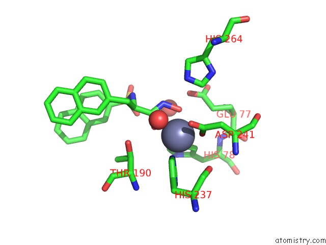
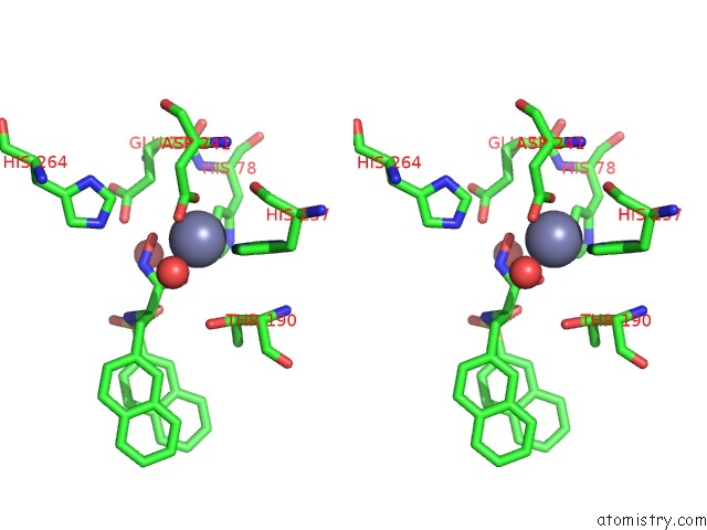
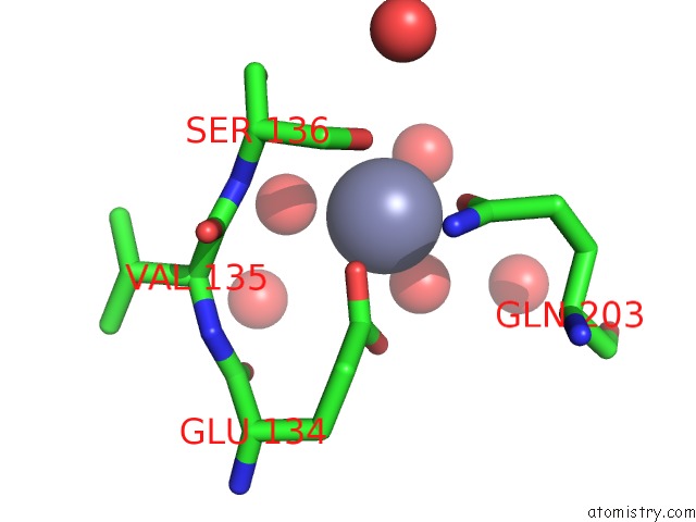
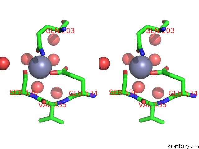
Zinc binding site 1 out of 7 in 2ves
Go back to
Zinc binding site 1 out
of 7 in the Crystal Structure of Lpxc From Pseudomonas Aeruginosa Complexed with the Potent Bb-78485 Inhibitor
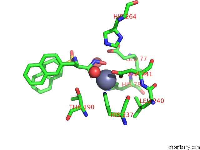
Mono view
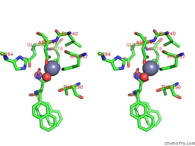
Stereo pair view
Mono view
Stereo pair view
A full contact list of Zinc with other atoms in the Zn binding
site number 1 of Crystal Structure of Lpxc From Pseudomonas Aeruginosa Complexed with the Potent Bb-78485 Inhibitor within 5.0Å range:
|
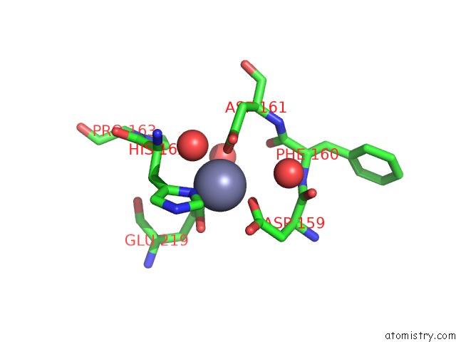
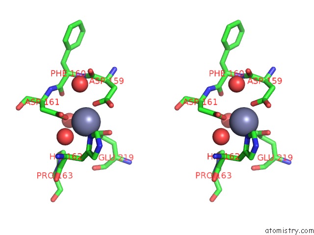
Zinc binding site 2 out of 7 in 2ves
Go back to
Zinc binding site 2 out
of 7 in the Crystal Structure of Lpxc From Pseudomonas Aeruginosa Complexed with the Potent Bb-78485 Inhibitor
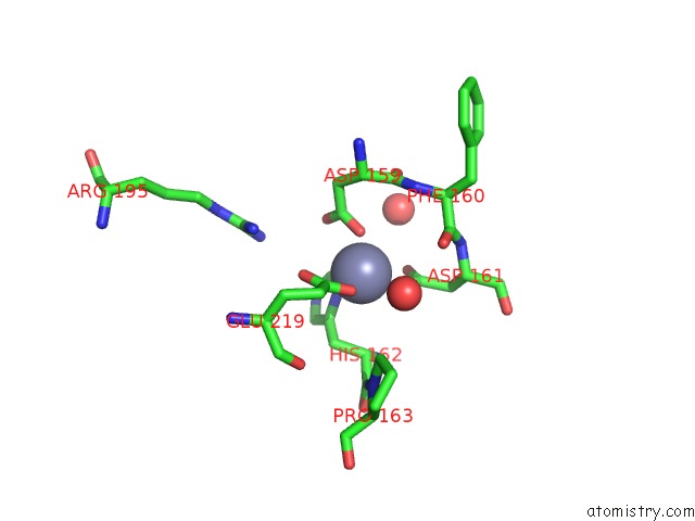
Mono view
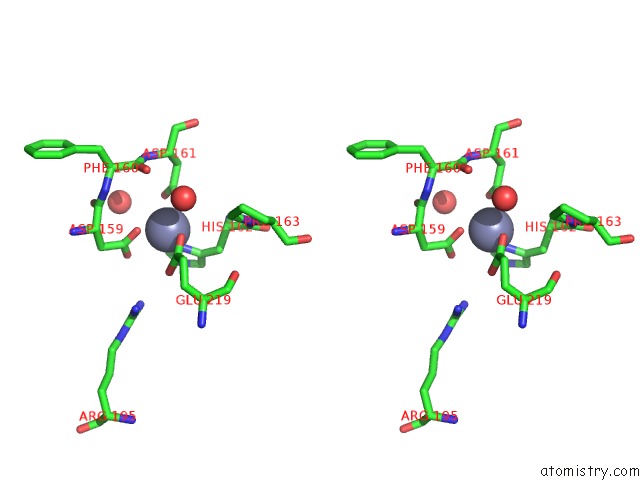
Stereo pair view
Mono view
Stereo pair view
A full contact list of Zinc with other atoms in the Zn binding
site number 2 of Crystal Structure of Lpxc From Pseudomonas Aeruginosa Complexed with the Potent Bb-78485 Inhibitor within 5.0Å range:
|
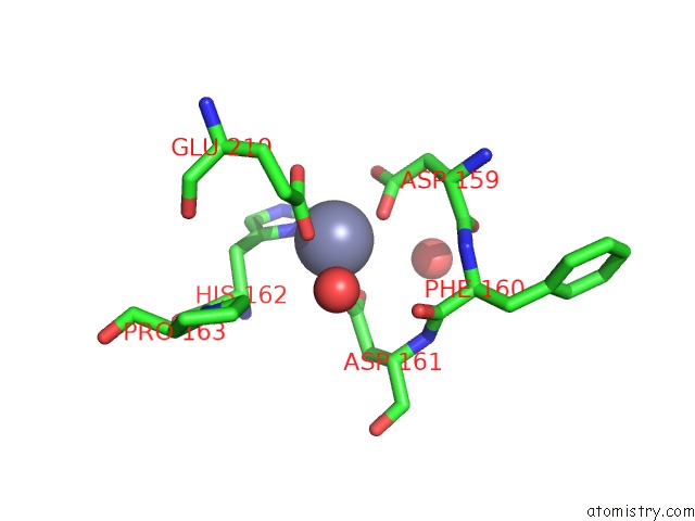
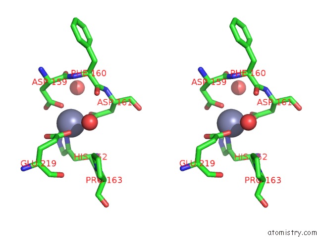
Zinc binding site 3 out of 7 in 2ves
Go back to
Zinc binding site 3 out
of 7 in the Crystal Structure of Lpxc From Pseudomonas Aeruginosa Complexed with the Potent Bb-78485 Inhibitor
Mono view
Stereo pair view
Mono view
Stereo pair view
A full contact list of Zinc with other atoms in the Zn binding
site number 3 of Crystal Structure of Lpxc From Pseudomonas Aeruginosa Complexed with the Potent Bb-78485 Inhibitor within 5.0Å range:
|
Zinc binding site 4 out of 7 in 2ves
Go back to
Zinc binding site 4 out
of 7 in the Crystal Structure of Lpxc From Pseudomonas Aeruginosa Complexed with the Potent Bb-78485 Inhibitor
Mono view
Stereo pair view
Mono view
Stereo pair view
A full contact list of Zinc with other atoms in the Zn binding
site number 4 of Crystal Structure of Lpxc From Pseudomonas Aeruginosa Complexed with the Potent Bb-78485 Inhibitor within 5.0Å range:
|
Zinc binding site 5 out of 7 in 2ves
Go back to
Zinc binding site 5 out
of 7 in the Crystal Structure of Lpxc From Pseudomonas Aeruginosa Complexed with the Potent Bb-78485 Inhibitor
Mono view
Stereo pair view
Mono view
Stereo pair view
A full contact list of Zinc with other atoms in the Zn binding
site number 5 of Crystal Structure of Lpxc From Pseudomonas Aeruginosa Complexed with the Potent Bb-78485 Inhibitor within 5.0Å range:
|
Zinc binding site 6 out of 7 in 2ves
Go back to
Zinc binding site 6 out
of 7 in the Crystal Structure of Lpxc From Pseudomonas Aeruginosa Complexed with the Potent Bb-78485 Inhibitor
Mono view
Stereo pair view
Mono view
Stereo pair view
A full contact list of Zinc with other atoms in the Zn binding
site number 6 of Crystal Structure of Lpxc From Pseudomonas Aeruginosa Complexed with the Potent Bb-78485 Inhibitor within 5.0Å range:
|
Zinc binding site 7 out of 7 in 2ves
Go back to
Zinc binding site 7 out
of 7 in the Crystal Structure of Lpxc From Pseudomonas Aeruginosa Complexed with the Potent Bb-78485 Inhibitor
Mono view
Stereo pair view
Mono view
Stereo pair view
A full contact list of Zinc with other atoms in the Zn binding
site number 7 of Crystal Structure of Lpxc From Pseudomonas Aeruginosa Complexed with the Potent Bb-78485 Inhibitor within 5.0Å range:
|
Reference:
I.Mochalkin,
J.D.Knafels,
S.Lightle.
Crystal Structure of Lpxc From Pseudomonas Aeruginosa Complexed with the Potent Bb-78485 Inhibitor. Protein Sci. V. 17 450 2008.
ISSN: ISSN 0961-8368
PubMed: 18287278
DOI: 10.1110/PS.073324108
Page generated: Thu Oct 17 04:16:20 2024
ISSN: ISSN 0961-8368
PubMed: 18287278
DOI: 10.1110/PS.073324108
Last articles
Zn in 9MJ5Zn in 9HNW
Zn in 9G0L
Zn in 9FNE
Zn in 9DZN
Zn in 9E0I
Zn in 9D32
Zn in 9DAK
Zn in 8ZXC
Zn in 8ZUF