Zinc »
PDB 2v20-2vh9 »
2v9l »
Zinc in PDB 2v9l: L-Rhamnulose-1-Phosphate Aldolase From Escherichia Coli (Mutant Q6Y- E192A)
Enzymatic activity of L-Rhamnulose-1-Phosphate Aldolase From Escherichia Coli (Mutant Q6Y- E192A)
All present enzymatic activity of L-Rhamnulose-1-Phosphate Aldolase From Escherichia Coli (Mutant Q6Y- E192A):
4.1.2.19;
4.1.2.19;
Protein crystallography data
The structure of L-Rhamnulose-1-Phosphate Aldolase From Escherichia Coli (Mutant Q6Y- E192A), PDB code: 2v9l
was solved by
D.Grueninger,
G.E.Schulz,
with X-Ray Crystallography technique. A brief refinement statistics is given in the table below:
| Resolution Low / High (Å) | 10.00 / 1.23 |
| Space group | P 4 21 2 |
| Cell size a, b, c (Å), α, β, γ (°) | 107.059, 107.059, 57.161, 90.00, 90.00, 90.00 |
| R / Rfree (%) | 9 / 12.2 |
Zinc Binding Sites:
The binding sites of Zinc atom in the L-Rhamnulose-1-Phosphate Aldolase From Escherichia Coli (Mutant Q6Y- E192A)
(pdb code 2v9l). This binding sites where shown within
5.0 Angstroms radius around Zinc atom.
In total only one binding site of Zinc was determined in the L-Rhamnulose-1-Phosphate Aldolase From Escherichia Coli (Mutant Q6Y- E192A), PDB code: 2v9l:
In total only one binding site of Zinc was determined in the L-Rhamnulose-1-Phosphate Aldolase From Escherichia Coli (Mutant Q6Y- E192A), PDB code: 2v9l:
Zinc binding site 1 out of 1 in 2v9l
Go back to
Zinc binding site 1 out
of 1 in the L-Rhamnulose-1-Phosphate Aldolase From Escherichia Coli (Mutant Q6Y- E192A)
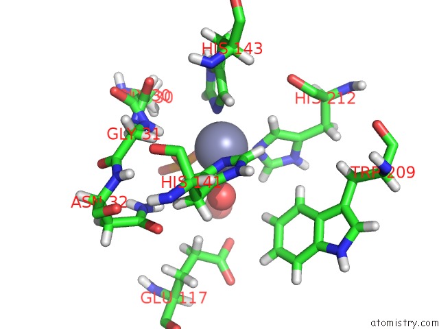
Mono view
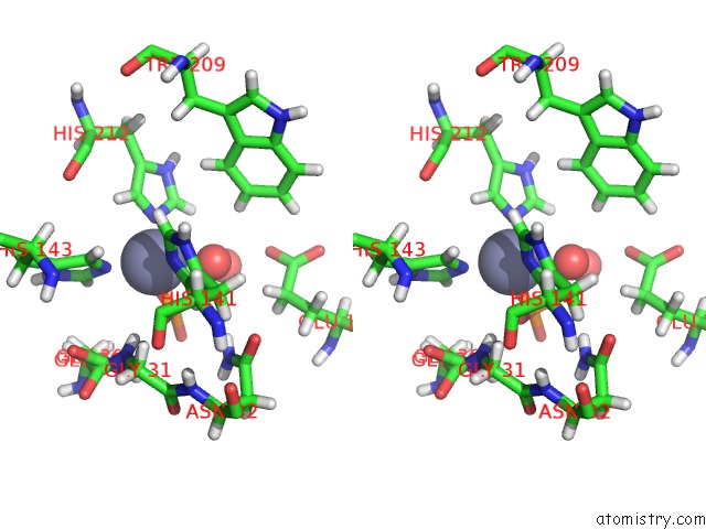
Stereo pair view
Mono view
Stereo pair view
A full contact list of Zinc with other atoms in the Zn binding
site number 1 of L-Rhamnulose-1-Phosphate Aldolase From Escherichia Coli (Mutant Q6Y- E192A) within 5.0Å range:
|
Reference:
D.Grueninger,
N.Treiber,
M.O.P.Ziegler,
J.W.A.Koetter,
M.-S.Schulze,
G.E.Schulz.
Designed Protein-Protein Association. Science V. 319 206 2008.
ISSN: ISSN 0036-8075
PubMed: 18187656
DOI: 10.1126/SCIENCE.1150421
Page generated: Thu Oct 17 04:14:25 2024
ISSN: ISSN 0036-8075
PubMed: 18187656
DOI: 10.1126/SCIENCE.1150421
Last articles
Zn in 9MJ5Zn in 9HNW
Zn in 9G0L
Zn in 9FNE
Zn in 9DZN
Zn in 9E0I
Zn in 9D32
Zn in 9DAK
Zn in 8ZXC
Zn in 8ZUF